
-
Product Name
BAG6 Polyclonal Antibody
- Documents
-
Description
Polyclonal antibody to BAG6
-
Tested applications
WB
-
Species reactivity
Human, Mouse, Rat
-
Alternative names
BAG6 antibody; BAG-6 antibody; BAT3 antibody; D6S52E antibody; G3 antibody; BCL2 associated athanogene 6 antibody
-
Isotype
Rabbit IgG
-
Preparation
Antigen: Recombinant fusion protein containing a sequence corresponding to amino acids 1-200 of human BAG6 (NP_001092004.1).
-
Clonality
Polyclonal
-
Formulation
PBS with 0.02% sodium azide, 50% glycerol, pH7.3.
-
Storage instructions
Store at -20℃. Avoid freeze / thaw cycles.
-
Applications
WB 1:500 - 1:2000
-
Validations
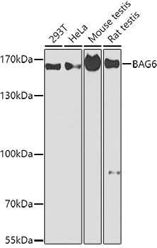
Western blot - BAG6 Polyclonal Antibody
Western blot analysis of extracts of various cell lines, using BAG6 antibody at 1:1000 dilution.Secondary antibody: HRP Goat Anti-Rabbit IgG (H+L) at 1:10000 dilution.Lysates/proteins: 25ug per lane.Blocking buffer: 3% nonfat dry milk in TBST.Detection: ECL Basic Kit .Exposure time: 90s.
-
Background
ATP-independent molecular chaperone preventing the aggregation of misfolded and hydrophobic patches-containing proteins. Functions as part of a cytosolic protein quality control complex, the BAG6/BAT3 complex, which maintains these client proteins in a soluble state and participates to their proper delivery to the endoplasmic reticulum or alternatively can promote their sorting to the proteasome where they undergo degradation. The BAG6/BAT3 complex is involved in the post-translational delivery of tail-anchored/type II transmembrane proteins to the endoplasmic reticulum membrane. Recruited to ribosomes, it interacts with the transmembrane region of newly synthesized tail-anchored proteins and together with SGTA and ASNA1 mediates their delivery to the endoplasmic reticulum. Client proteins that cannot be properly delivered to the endoplasmic reticulum are ubiquitinated by RNF126, an E3 ubiquitin-protein ligase associated with BAG6 and are sorted to the proteasome. SGTA which prevents the recruitment of RNF126 to BAG6 may negatively regulate the ubiquitination and the proteasomal degradation of client proteins. Similarly, the BAG6/BAT3 complex also functions as a sorting platform for proteins of the secretory pathway that are mislocalized to the cytosol either delivering them to the proteasome for degradation or to the endoplasmic reticulum. The BAG6/BAT3 complex also plays a role in the endoplasmic reticulum-associated degradation (ERAD), a quality control mechanism that eliminates unwanted proteins of the endoplasmic reticulum through their retrotranslocation to the cytosol and their targeting to the proteasome. It maintains these retrotranslocated proteins in an unfolded yet soluble state condition in the cytosol to ensure their proper delivery to the proteasome. BAG6 is also required for selective ubiquitin-mediated degradation of defective nascent chain polypeptides by the proteasome. In this context, it may participate to the production of antigenic peptides and play a role in antigen presentation in immune response (By similarity). BAG6 is also involved in endoplasmic reticulum stress-induced pre-emptive quality control, a mechanism that selectively attenuates the translocation of newly synthesized proteins into the endoplasmic reticulum and reroutes them to the cytosol for proteasomal degradation. BAG6 may ensure the proper degradation of these proteins and thereby protects the endoplasmic reticulum from protein overload upon stress. By inhibiting the polyubiquitination and subsequent proteasomal degradation of HSPA2 it may also play a role in the assembly of the synaptonemal complex during spermatogenesis (By similarity). Also positively regulates apoptosis by interacting with and stabilizing the proapoptotic factor AIFM1 (By similarity). By controlling the steady-state expression of the IGF1R receptor, indirectly regulates the insulin-like growth factor receptor signaling pathway.; Involved in DNA damage-induced apoptosis: following DNA damage, accumulates in the nucleus and forms a complex with p300/EP300, enhancing p300/EP300-mediated p53/TP53 acetylation leading to increase p53/TP53 transcriptional activity. When nuclear, may also act as a component of some chromatin regulator complex that regulates histone 3 'Lys-4' dimethylation (H3K4me2).; Released extracellularly via exosomes, it is a ligand of the natural killer/NK cells receptor NCR3 and stimulates NK cells cytotoxicity. It may thereby trigger NK cells cytotoxicity against neighboring tumor cells and immature myeloid dendritic cells (DC).; Mediates ricin-induced apoptosis.
Related Products / Services
Please note: All products are "FOR RESEARCH USE ONLY AND ARE NOT INTENDED FOR DIAGNOSTIC OR THERAPEUTIC USE"