
-
Product Name
Anti-WDR70 antibody
- Documents
-
Description
Rabbit polyclonal antibody to WDR70
-
Tested applications
WB, FC
-
Species reactivity
Human
-
Isotype
Rabbit IgG
-
Preparation
This antigen of this antibody was synthetic peptide within human wdr70 aa 1-50.
-
Clonality
Polyclonal
-
Formulation
Liquid, 1*PBS (pH7.4), 0.2% BSA, 50% Glycerol. Preservative: 0.05% Sodium Azide.
-
Storage instructions
Store at +4℃ after thawing. Aliquot store at -20℃. Avoid repeated freeze / thaw cycles.
-
Applications
WB:1:500-1:1,000
FC:1:50-1:100
-
Validations
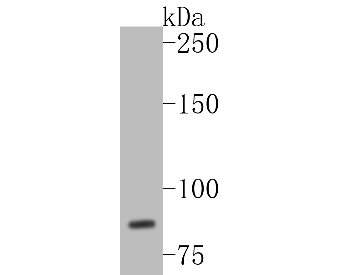
Fig1:; Western blot analysis of WDR70 on Hela cell lysates. Proteins were transferred to a PVDF membrane and blocked with 5% BSA in PBS for 1 hour at room temperature. The primary antibody ( 1/500) was used in 5% BSA at room temperature for 2 hours. Goat Anti-Rabbit IgG - HRP Secondary Antibody (HA1001) at 1:5,000 dilution was used for 1 hour at room temperature.
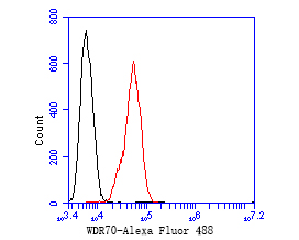
Fig2:; Flow cytometric analysis of WDR70 was done on SiHa cells. The cells were fixed, permeabilized and stained with the primary antibody ( 1/50) (red). After incubation of the primary antibody at room temperature for an hour, the cells were stained with a Alexa Fluor 488-conjugated Goat anti-Rabbit IgG Secondary antibody at 1/1000 dilution for 30 minutes.Unlabelled sample was used as a control (cells without incubation with primary antibody; black).
- Background
-
References
- Zeng M. et. al. CRL4(Wdr70) regulates H2B monoubiquitination and facilitates Exo1-dependent resection. Nat Commun. 2016 Apr
- Zeng M. et. al. Wdr70 regulates histone modification and genomic maintenance in fission yeast. Biochim Biophys Acta Mol Cell Res. 2020 May
Related Products / Services
Please note: All products are "FOR RESEARCH USE ONLY AND ARE NOT INTENDED FOR DIAGNOSTIC OR THERAPEUTIC USE"