
-
Product Name
Anti-WASF2 antibody
- Documents
-
Description
Rabbit monoclonal antibody to WASF2
-
Tested applications
WB, IHC-P
-
Species reactivity
Human, Mouse, Rat
-
Alternative names
IMD2 antibody; SCAR2 antibody; WASF4 antibody; WAVE2 antibody; dJ393P12.2 antibody
-
Isotype
Rabbit IgG
-
Preparation
This antigen of this antibody was synthetic peptide within human wasf2 aa 440-480.
-
Clonality
Monoclonal
-
Formulation
Liquid, 1*TBS (pH7.4), 0.05% BSA, 40% Glycerol. Preservative: 0.05% Sodium Azide.
-
Storage instructions
Store at +4℃ after thawing. Aliquot store at -20℃ or -80℃. Avoid repeated freeze / thaw cycles.
-
Applications
WB: 1:500-1:2,000
IHC-P: 1:50-1:200
-
Validations
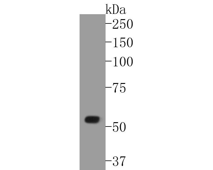
Fig1:; Western blot analysis of WASF2 on human placenta tissue lysates. Proteins were transferred to a PVDF membrane and blocked with 5% BSA in PBS for 1 hour at room temperature. The primary antibody ( 1/500) was used in 5% BSA at room temperature for 2 hours. Goat Anti-Rabbit IgG - HRP Secondary Antibody (HA1001) at 1:5,000 dilution was used for 1 hour at room temperature.
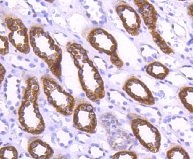
Fig2:; Immunohistochemical analysis of paraffin-embedded human kidney tissue using anti-WASF2 antibody. The section was pre-treated using heat mediated antigen retrieval with Tris-EDTA buffer (pH 8.0-8.4) for 20 minutes.The tissues were blocked in 5% BSA for 30 minutes at room temperature, washed with ddH; 2; O and PBS, and then probed with the primary antibody ( 1/50) for 30 minutes at room temperature. The detection was performed using an HRP conjugated compact polymer system. DAB was used as the chromogen. Tissues were counterstained with hematoxylin and mounted with DPX.
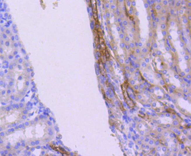
Fig3:; Immunohistochemical analysis of paraffin-embedded mouse kidney tissue using anti-WASF2 antibody. The section was pre-treated using heat mediated antigen retrieval with Tris-EDTA buffer (pH 8.0-8.4) for 20 minutes.The tissues were blocked in 5% BSA for 30 minutes at room temperature, washed with ddH; 2; O and PBS, and then probed with the primary antibody ( 1/50) for 30 minutes at room temperature. The detection was performed using an HRP conjugated compact polymer system. DAB was used as the chromogen. Tissues were counterstained with hematoxylin and mounted with DPX.
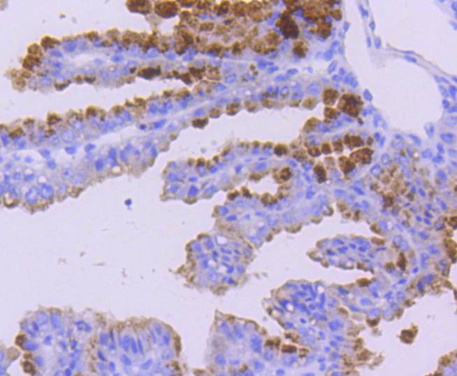
Fig4:; Immunohistochemical analysis of paraffin-embedded mouse placenta tissue using anti-WASF2 antibody. The section was pre-treated using heat mediated antigen retrieval with Tris-EDTA buffer (pH 8.0-8.4) for 20 minutes.The tissues were blocked in 5% BSA for 30 minutes at room temperature, washed with ddH; 2; O and PBS, and then probed with the primary antibody ( 1/50) for 30 minutes at room temperature. The detection was performed using an HRP conjugated compact polymer system. DAB was used as the chromogen. Tissues were counterstained with hematoxylin and mounted with DPX.
- Background
-
References
- Jia, S. et al. 2014. Down-regulation of WAVE2, WASP family verprolin-homologous protein 2, in gastric cancer indicates lymph node metastasis and cell migration. Anticancer research. 34: 2185-94.
- Goh, WI. et al. 2012. mDia1 and WAVE2 proteins interact directly with IRSp53 in filopodia and are involved in filopodium formation. J. Biol. Chem. 287: 4702-4714.
Related Products / Services
Please note: All products are "FOR RESEARCH USE ONLY AND ARE NOT INTENDED FOR DIAGNOSTIC OR THERAPEUTIC USE"