
-
Product Name
Anti-TUBA8 antibody
- Documents
-
Description
Mouse monoclonal antibody to TUBA8
-
Tested applications
WB, ICC, IHC-P, FC
-
Species reactivity
Human, Rat
-
Alternative names
CDCBM8 antibody; TUBAL2 antibody
-
Isotype
Mouse IgG2b
-
Preparation
This antigen of this antibody was recombinant protein
-
Clonality
Monoclonal
-
Formulation
Liquid, 1*TBS (pH7.4), 1%BSA, Preservative: 0.05% Sodium Azide.
-
Storage instructions
Store at +4℃after thawing. Aliquot store at -20℃ or -80℃. Avoid repeated freeze / thaw cycles.
-
Applications
WB: 1:500-1:2,000
ICC: 1:100-1:500
IHC-P: 1:100-1:500
FC: 1:100-1:200
-
Validations
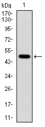
Fig1: Western blot analysis of TUBA8 on human TUBA8 recombinant protein using anti-TUBA8 antibody at 1/1,000 dilution.
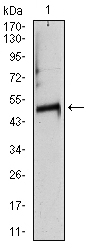
Fig2: Western blot analysis of TUBA8 on at heart tissue lysate using anti-TUBA8 antibody at 1/1,000 dilution.
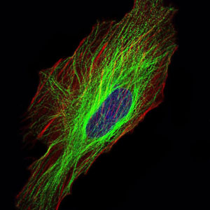
Fig3: ICC staining TUBA8 (green) and actin filaments (red) in Hela cells. The nuclear counter stain is DAPI (blue). Cells were fixed in paraformaldehyde, permeabilised with 0.25% Triton X100/PBS.
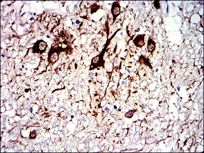
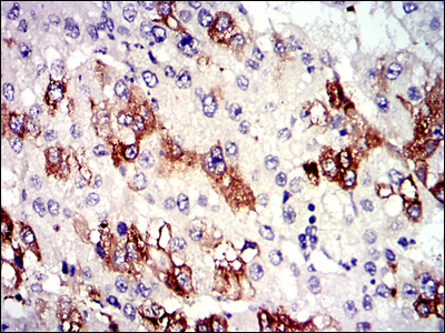
Fig4: Immunohistochemical analysis of paraffin-embedded medulla oblongata tissues using anti-TUBA8 antibody. Counter stained with hematoxylin.
Fig5: Immunohistochemical analysis of paraffin-embedded liver cancer tissues using anti-TUBA8 antibody. Counter stained with hematoxylin.
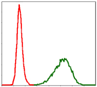
Fig6: Flow cytometric analysis of NIH/3T3 cells with TUBA8 antibody at 1/100 dilution (green) compared with an unlabelled control (cells without incubation with primary antibody; red).
- Background
-
References
- Ehnert S et al. TGF-β1 impairs mechanosensation of human osteoblasts via HDAC6-mediated shortening and distortion of primary cilia. Journal of molecular medicine (Berlin, Germany). 95: 653-663 (2017).
- Noack K et al. Analysis of the interplay between all-trans retinoic acid and histone deacetylase inhibitors in leukemic cells. Archives of toxicology. 91: 2191-2208 (2017).
Related Products / Services
Please note: All products are "FOR RESEARCH USE ONLY AND ARE NOT INTENDED FOR DIAGNOSTIC OR THERAPEUTIC USE"