
-
Product Name
Anti-TMEM177 antibody
- Documents
-
Description
Mouse monoclonal antibody to TMEM177
-
Tested applications
WB, ICC, IHC-P
-
Species reactivity
Human
-
Isotype
Mouse IgG1
-
Preparation
This antigen of this antibody was recombinant protein
-
Clonality
Monoclonal
-
Formulation
Liquid, 1*PBS (pH7.4), 0.2% BSA, 40% Glycerol. Preservative: 0.05% Sodium Azide.
-
Storage instructions
Store at +4℃ after thawing. Aliquot store at -20℃ or -80℃. Avoid repeated freeze / thaw cycles
-
Applications
WB: 1:500-1:1,000
ICC: 1:50-1:200
IHC-P: 1:50-1:200
-
Validations
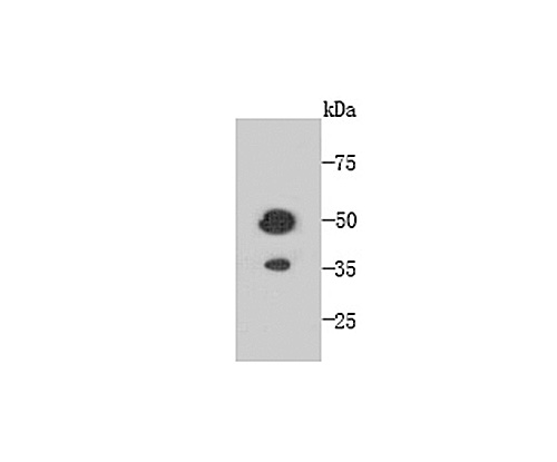
Fig1: Western blot analysis of TMEM177 on recombinant protein using anti-TMEM177 antibody at 1/1,000 dilution.
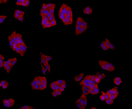
Fig2: ICC staining TMEM177 in Hela cells (red). The nuclear counter stain is DAPI (blue). Cells were fixed in paraformaldehyde, permeabilised with 0.25% Triton X100/PBS.
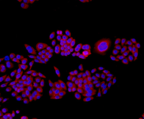
Fig3: ICC staining TMEM177 in HepG2 cells (red). The nuclear counter stain is DAPI (blue). Cells were fixed in paraformaldehyde, permeabilised with 0.25% Triton X100/PBS.
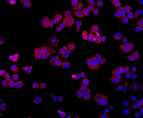
Fig4: ICC staining TMEM177 in MCF-7 cells (red). The nuclear counter stain is DAPI (blue). Cells were fixed in paraformaldehyde, permeabilised with 0.25% Triton X100/PBS.
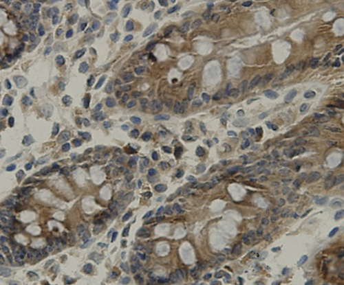
Fig5: Immunohistochemical analysis of paraffin-embedded human colon tissue using anti-TMEM177 antibody. Counter stained with hematoxylin.
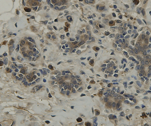
Fig6: Immunohistochemical analysis of paraffin-embedded human breast tissue using anti-TMEM177 antibody. Counter stained with hematoxylin.
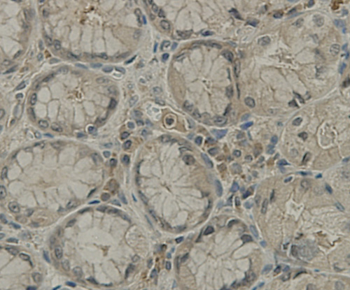
Fig7: Immunohistochemical analysis of paraffin-embedded hman stamoch tissue using anti-TMEM177 antibody. Counter stained with hematoxylin.
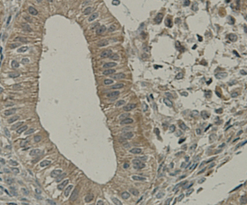
Fig8: Immunohistochemical analysis of paraffin-embedded human uterus muscle tissue using anti-TMEM177 antibody. Counter stained with hematoxylin.
- Background
-
References
- Hopf TA et al. Three-dimensional structures of membrane proteins from genomic sequencing. Cell 149(7):1607-21 (2012).
- Bracey MH et al. Structural adaptations in a membrane enzyme that terminates endocannabinoid.
Related Products / Services
Please note: All products are "FOR RESEARCH USE ONLY AND ARE NOT INTENDED FOR DIAGNOSTIC OR THERAPEUTIC USE"