
-
Product Name
Anti-SPATA5L1 antibody
- Documents
-
Description
Mouse monoclonal antibody to SPATA5L1
-
Tested applications
WB, ICC, IHC-P, FC
-
Species reactivity
Human, Rat
-
Isotype
Mouse IgG1
-
Preparation
This antigen of this antibody was synthetic peptide within human aa 350-600.
-
Clonality
Monoclonal
-
Formulation
Liquid, 1*PBS (pH7.4), 0.2% BSA, 50% Glycerol. Preservative: 0.05% Sodium Azide.
-
Storage instructions
Store at +4℃ after thawing. Aliquot store at -20℃. Avoid repeated freeze / thaw cycles.
-
Applications
WB:1:500-1:2,000
ICC:1:50-1:200
IHC-P:1:50-1:200
FC:1:50-1:100
-
Validations
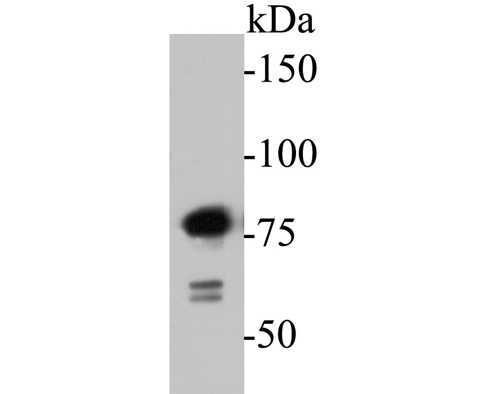
Fig1: Western blot analysis of SPATA5L1 on K562 cell lysates. Proteins were transferred to a PVDF membrane and blocked with 5% BSA in PBS for 1 hour at room temperature. The primary antibody ( 1/500) was used in 5% BSA at room temperature for 2 hours. Goat Anti-Rabbit IgG - HRP Secondary Antibody (HA1001) at 1:5,000 dilution was used for 1 hour at room temperature.
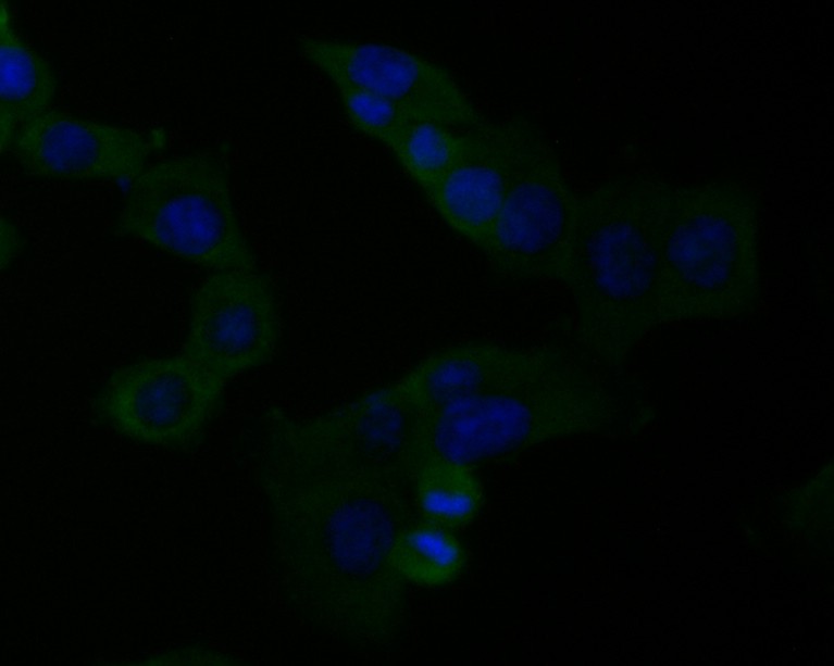
Fig2: ICC staining of SPATA5L1 in HCT116 cells (green). Formalin fixed cells were permeabilized with 0.1% Triton X-100 in TBS for 10 minutes at room temperature and blocked with 1% Blocker BSA for 15 minutes at room temperature. Cells were probed with the primary antibody ( 1/50) for 1 hour at room temperature, washed with PBS. Alexa Fluor®488 Goat anti-Mouse IgG was used as the secondary antibody at 1/1,000 dilution. The nuclear counter stain is DAPI (blue).
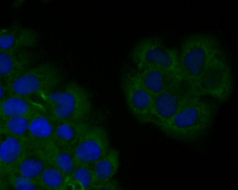
Fig3: ICC staining of SPATA5L1 in JAR cells (green). Formalin fixed cells were permeabilized with 0.1% Triton X-100 in TBS for 10 minutes at room temperature and blocked with 1% Blocker BSA for 15 minutes at room temperature. Cells were probed with the primary antibody ( 1/50) for 1 hour at room temperature, washed with PBS. Alexa Fluor®488 Goat anti-Mouse IgG was used as the secondary antibody at 1/1,000 dilution. The nuclear counter stain is DAPI (blue).
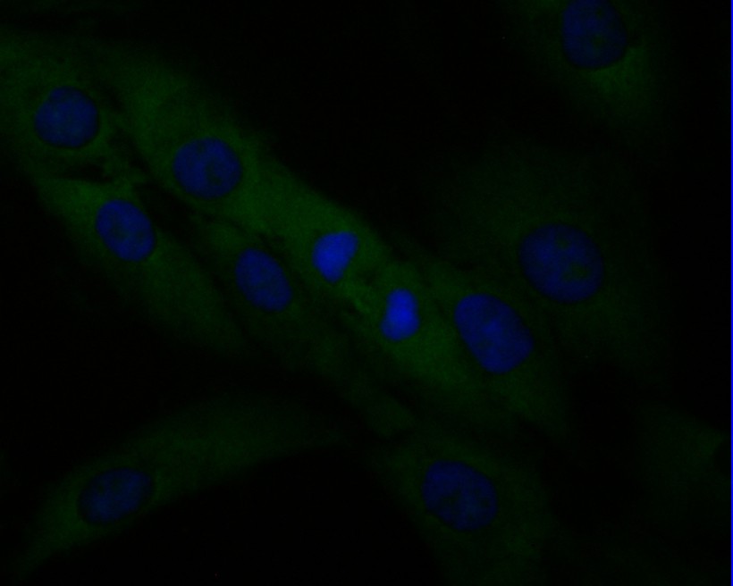
Fig4: ICC staining of SPATA5L1 in MG-63 cells (green). Formalin fixed cells were permeabilized with 0.1% Triton X-100 in TBS for 10 minutes at room temperature and blocked with 1% Blocker BSA for 15 minutes at room temperature. Cells were probed with the primary antibody ( 1/50) for 1 hour at room temperature, washed with PBS. Alexa Fluor®488 Goat anti-Mouse IgG was used as the secondary antibody at 1/1,000 dilution. The nuclear counter stain is DAPI (blue).
Fig5: Immunohistochemical analysis of paraffin-embedded rat seminal vesicle tissue using anti-SPATA5L1 antibody. The section was pre-treated using heat mediated antigen retrieval with Tris-EDTA buffer (pH 8.0-8.4) for 20 minutes.The tissues were blocked in 5% BSA for 30 minutes at room temperature, washed with ddH2O and PBS, and then probed with the primary antibody ( 1/50) for 30 minutes at room temperature. The detection was performed using an HRP conjugated compact polymer system. DAB was used as the chromogen. Tissues were counterstained with hematoxylin and mounted with DPX.
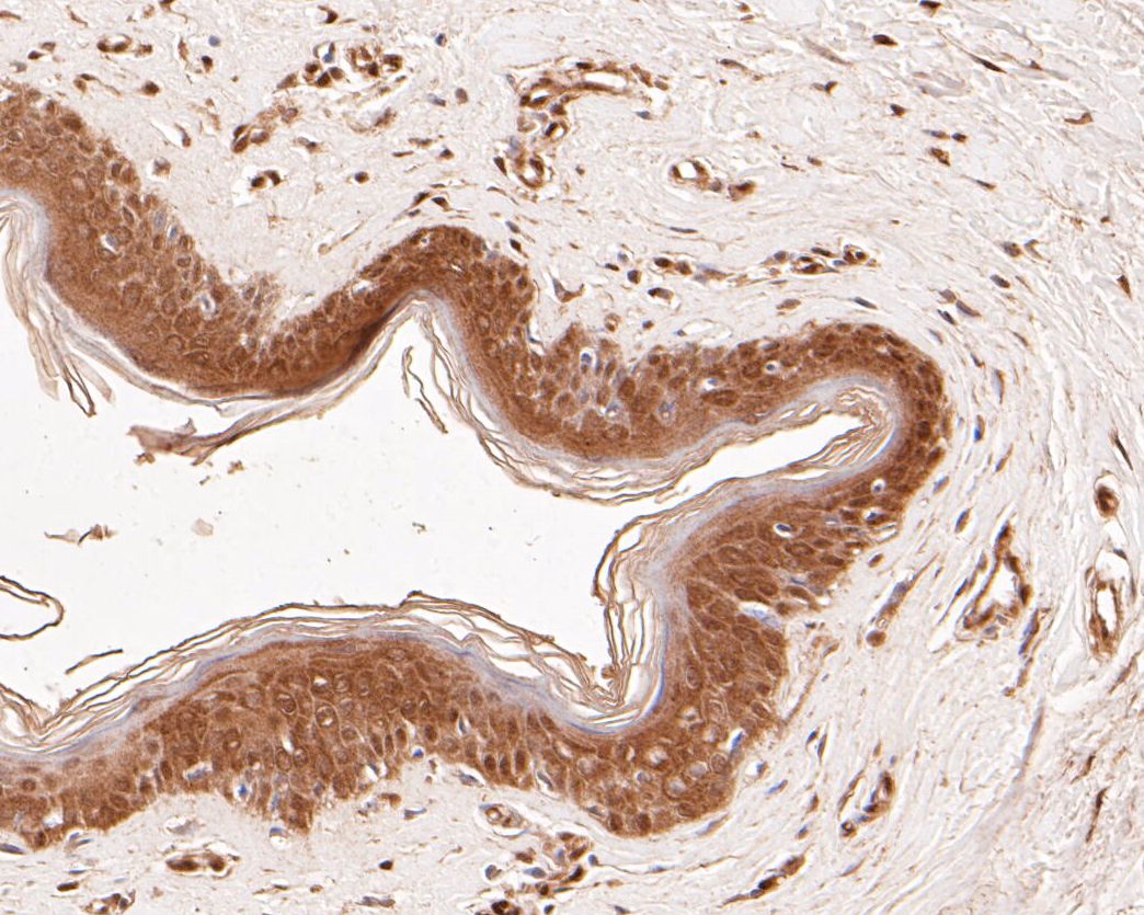
Fig6: Immunohistochemical analysis of paraffin-embedded human skin tissue using anti-SPATA5L1 antibody. The section was pre-treated using heat mediated antigen retrieval with Tris-EDTA buffer (pH 8.0-8.4) for 20 minutes.The tissues were blocked in 5% BSA for 30 minutes at room temperature, washed with ddH2O and PBS, and then probed with the primary antibody ( 1/50) for 30 minutes at room temperature. The detection was performed using an HRP conjugated compact polymer system. DAB was used as the chromogen. Tissues were counterstained with hematoxylin and mounted with DPX.
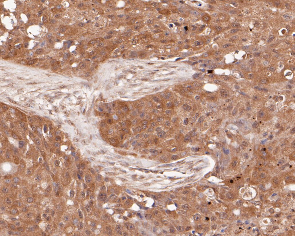
Fig7: Immunohistochemical analysis of paraffin-embedded human breast carcinoma tissue using anti-SPATA5L1 antibody. The section was pre-treated using heat mediated antigen retrieval with Tris-EDTA buffer (pH 8.0-8.4) for 20 minutes.The tissues were blocke
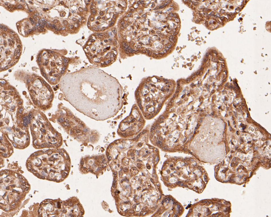
Fig8: Immunohistochemical analysis of paraffin-embedded human placenta tissue using anti-SPATA5L1 antibody. The section was pre-treated using heat mediated antigen retrieval with Tris-EDTA buffer (pH 8.0-8.4) for 20 minutes.The tissues were blocked in 5%
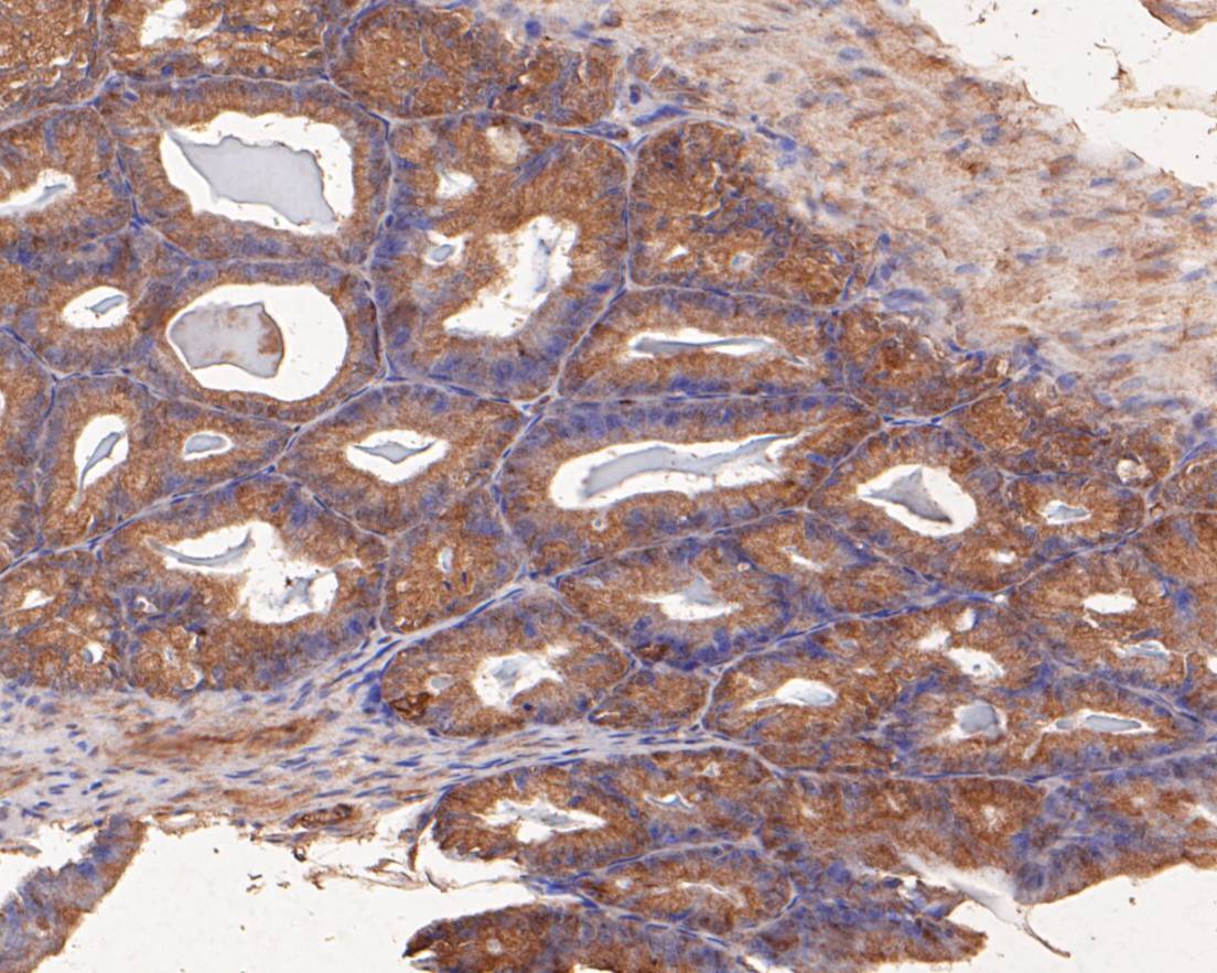
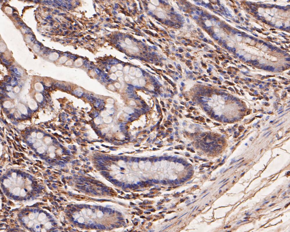
Fig9: Immunohistochemical analysis of paraffin-embedded human small intestine tissue using anti-SPATA5L1 antibody. The section was pre-treated using heat mediated antigen retrieval with Tris-EDTA buffer (pH 8.0-8.4) for 20 minutes.The tissues were blocked
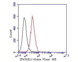
Fig10: Flow cytometric analysis of SPATA5L1 was done on JAR cells. The cells were fixed, permeabilized and stained with the primary antibody ( 1/50) (red). After incubation of the primary antibody at room temperature for an hour, the cells were
- Background
-
References
- Kubo Y. et. al. Association between kidney function and genetic polymorphisms in atherosclerotic and chronic kidney diseases: A cross-sectional study in Japanese male workers. PLoS One. 2017 Oct 10;12(10)
- Park H. et. al. A family-based association study after genome-wide linkage analysis identified two genetic loci for renal function in a Mongolian population. Kidney Int. 2013 Feb;83(2)
Related Products / Services
Please note: All products are "FOR RESEARCH USE ONLY AND ARE NOT INTENDED FOR DIAGNOSTIC OR THERAPEUTIC USE"