


-
Product Name
Anti-SMC3 antibody
- Documents
-
Description
Rabbit monoclonal antibody to SMC3
-
Tested applications
WB, ICC/IF, IHC-P, FC
-
Species reactivity
Human, Mouse, Rat
-
Alternative names
BAM antibody; BMH antibody; HCAP antibody; CDLS3 antibody; CSPG6 antibody; SMC3L1 antibody
-
Isotype
Rabbit IgG
-
Preparation
This antigen of this antibody was recombinant protein
-
Clonality
Monoclonal
-
Formulation
Liquid, 1*TBS (pH7.4), 0.05% BSA, 40% Glycerol. Preservative: 0.05% Sodium Azide.
-
Storage instructions
Store at +4℃ after thawing. Aliquot store at -20℃ or -80℃. Avoid repeated freeze / thaw cycles.
-
Applications
WB: 1:1,000-1:5,000
ICC/IF: 1:100-1:500
IHC-P: 1:50-1:200
FC: 1:50-1:100
-
Validations
Fig1: Western blot analysis of SMC3 on different cells lysates using anti-SMC3 antibody at 1/1,000 dilution.; Positive control:; Line1: HepG2; Line2: NIH-3T3; Line3:PC12
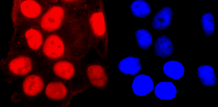
Fig2: ICC staining SMC3 in Hela cells (red). The nuclear counter stain is DAPI (blue). Cells were fixed in paraformaldehyde, permeabilised with 0.25% Triton X100/PBS.
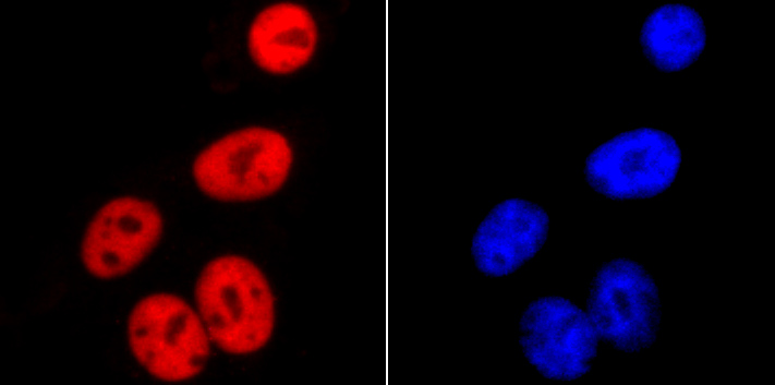
Fig3: ICC staining SMC3 in HepG2 cells (red). The nuclear counter stain is DAPI (blue). Cells were fixed in paraformaldehyde, permeabilised with 0.25% Triton X100/PBS.
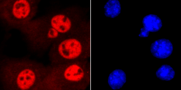
Fig4: ICC staining SMC3 in NIH-3T3 cells (red). The nuclear counter stain is DAPI (blue). Cells were fixed in paraformaldehyde, permeabilised with 0.25% Triton X100/PBS.
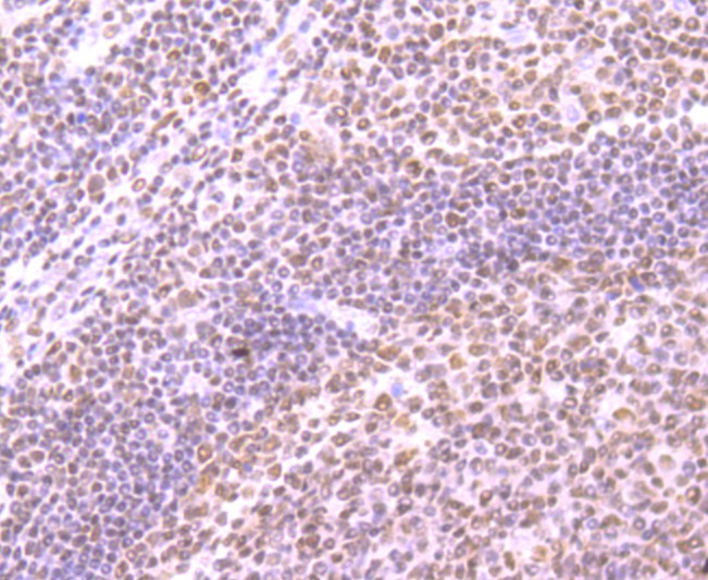
Fig5: Immunohistochemical analysis of paraffin-embedded human tonsil tissue using anti-SMC3 antibody. Counter stained with hematoxylin.
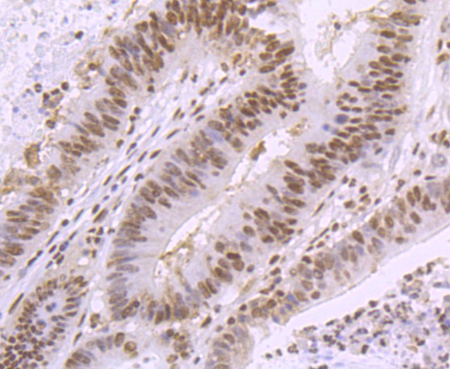
Fig6: Immunohistochemical analysis of paraffin-embedded human colon cancer tissue using anti-SMC3 antibody. Counter stained with hematoxylin.
Fig7: Immunohistochemical analysis of paraffin-embedded mouse colon tissue using anti-SMC3 antibody. Counter stained with hematoxylin.
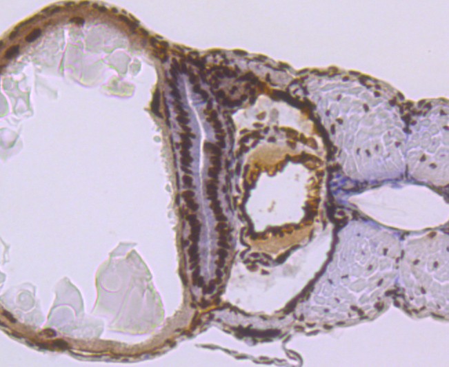
Fig8: Immunohistochemical analysis of paraffin-embedded zebrafish tissue using anti-SMC3 antibody. Counter stained with hematoxylin.
Fig9: Flow cytometric analysis of Hela cells with SMC3 antibody at 1/50 dilution (red) compared with an unlabelled control (cells without incubation with primary antibody; black). Alexa Fluor 488-conjugated goat anti rabbit IgG was used as the secondary a
- Background
-
References
- Kracker S et al. An inherited immunoglobulin class-switch recombination deficiency associated with a defect in the INO80 chromatin remodeling complex. J Allergy Clin Immunol 135:998-1007.e6 (2015).
- Qiu Z et al. Functional interactions between NURF and Ctcf regulate gene expression. Mol Cell Biol 35:224-37 (2015).
Related Products / Services
Please note: All products are "FOR RESEARCH USE ONLY AND ARE NOT INTENDED FOR DIAGNOSTIC OR THERAPEUTIC USE"