
-
Product Name
Anti-she antibody
- Documents
-
Description
Rabbit polyclonal antibody to she
-
Tested applications
WB, IF, IHC-P
-
Species reactivity
Zebrafish
-
Isotype
Rabbit IgG
-
Preparation
This antigen of this antibody was recombinant protein within zebrafish she aa 80-280.
-
Clonality
Polyclonal
-
Formulation
Liquid, 1*PBS (pH7.4), 0.2% BSA, 50% Glycerol. Preservative: 0.05% Sodium Azide.
-
Storage instructions
Store at +4℃ after thawing. Aliquot store at -20℃. Avoid repeated freeze / thaw cycles.
-
Applications
WB: 1:500-1:1000
IF: 1:50-1:200
IHC-P: 1:50-1:200
-
Validations
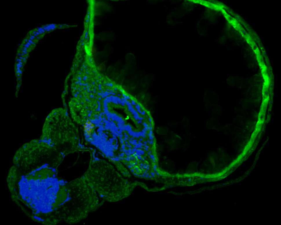
Fig1:; Immunofluorescence staining of paraffin- embedded Zebrafish using anti-SHE rabbit polyclonal antibody.The section was pre-treated using heat mediated antigen retrieval with Tris-EDTA buffer (pH 9.0) for 20 minutes. The tissues were blocked in 10% negative goat serum for 1 hour at room temperature, washed with PBS, and then probed with the antibody at 1/50 dilution for 10 hours at 4℃ and detected using Alexa Fluor™ 488 conjugate-Goat anti-Rabbit IgG (H+L) Secondary Antibody at a dilution of 1:500 for 1 hour at room temperature.
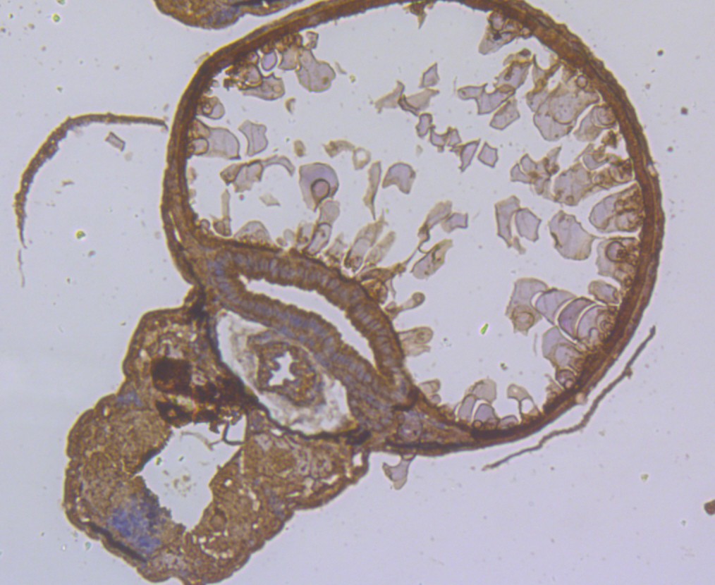
Fig2:; Immunohistochemical analysis of paraffin-embedded Zebrafish tissue using anti-SHE antibody. The section was pre-treated using heat mediated antigen retrieval with Tris-EDTA buffer (pH 8.0-8.4) for 20 minutes.The tissues were blocked in 5% BSA for 30 minutes at room temperature, washed with ddH; 2; O and PBS, and then probed with the antibody at 1/200 dilution, for 30 minutes at room temperature and detected using an HRP conjugated compact polymer system. DAB was used as the chrogen. Counter stained with hematoxylin and mounted with DPX.
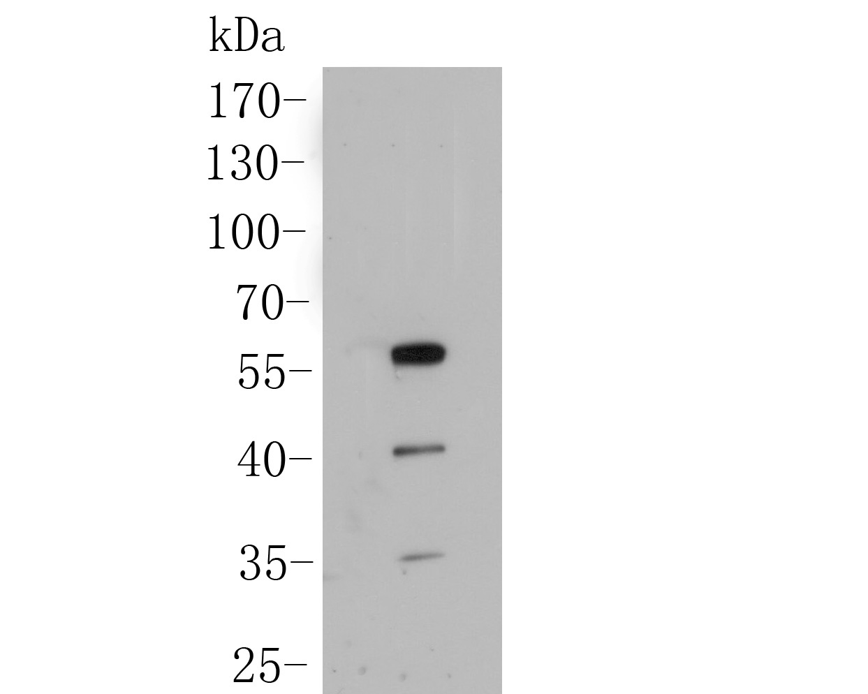
Fig3:; Western blot analysis of SHE on Zebrafish tissue lysate. Proteins were transferred to a PVDF membrane and blocked with 5% BSA in PBS for 1 hour at room temperature. The primary antibody ( 1/500) was used in 5% BSA at room temperature for 2 hours. Goat Anti-Rabbit IgG - HRP Secondary Antibody (HA1001) at 1:5,000 dilution was used for 1 hour at room temperature.
- Background
-
References
- Gaudet P.et.al.Phylogenetic-based propagation of functional annotations within the Gene Ontology consortium.Brief. Bioinformatics 12:449-462(2011).
Related Products / Services
Please note: All products are "FOR RESEARCH USE ONLY AND ARE NOT INTENDED FOR DIAGNOSTIC OR THERAPEUTIC USE"