
-
Product Name
Anti-PHF8 antibody
- Documents
-
Description
Rabbit polyclonal antibody to PHF8
-
Tested applications
WB, ICC, IHC-P, FC
-
Species reactivity
Human, Mouse, Rat
-
Alternative names
KDM7B antibody; JHDM1F antibody; MRXSSD antibody; ZNF422 antibody
-
Isotype
Rabbit IgG
-
Preparation
This antigen of this antibody was recombinant protein within human phf8 aa 150-350.
-
Clonality
Polyclonal
-
Formulation
Liquid, 1*PBS (pH7.4), 0.2% BSA, 50% Glycerol. Preservative: 0.05% Sodium Azide.
-
Storage instructions
Store at +4℃ after thawing. Aliquot store at -20℃. Avoid repeated freeze / thaw cycles.
-
Applications
WB: 1:500-1:2,000
ICC: 1:50-1:200
IHC-P: 1:50-1:200
FC: 1:50-1:100
-
Validations
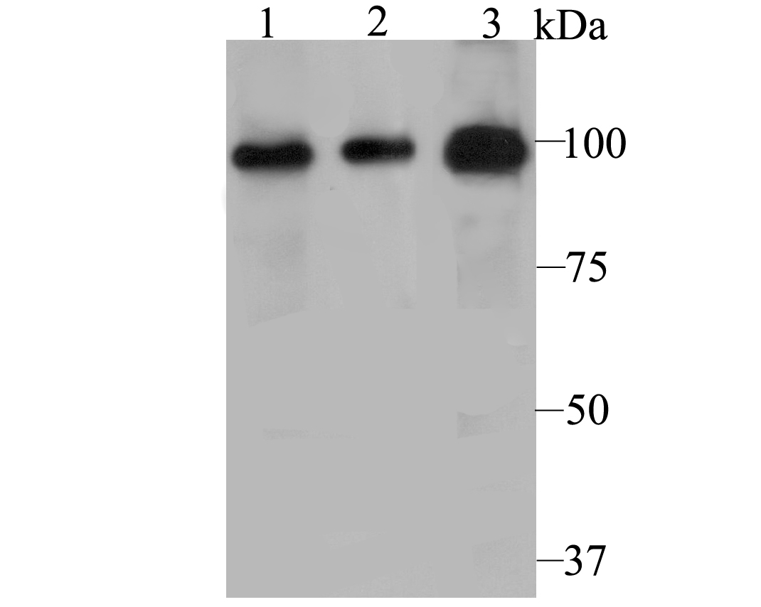
Fig1: Western blot analysis of PHF8 on different cell lysate using anti-PHF8 antibody at 1/500 dilution.; Positive control:; Lane 1: PC-3M; Lane 2: A431; Lane 3: Human kidney tissue
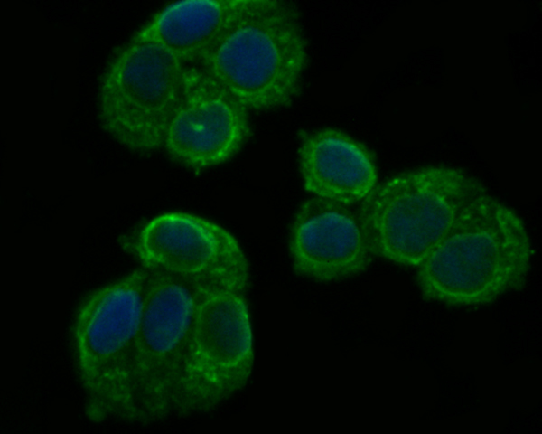
Fig2: ICC staining PHF8 in Hela cells (green). The nuclear counter stain is DAPI (blue). Cells were fixed in paraformaldehyde, permeabilised with 0.25% Triton X100/PBS.
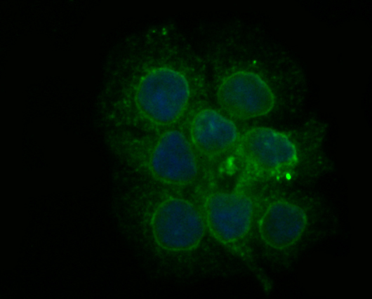
Fig3: ICC staining PHF8 in JAR cells (green). The nuclear counter stain is DAPI (blue). Cells were fixed in paraformaldehyde, permeabilised with 0.25% Triton X100/PBS.
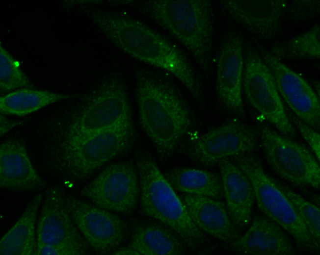
Fig4: ICC staining PHF8 in SiHa cells (green). The nuclear counter stain is DAPI (blue). Cells were fixed in paraformaldehyde, permeabilised with 0.25% Triton X100/PBS.
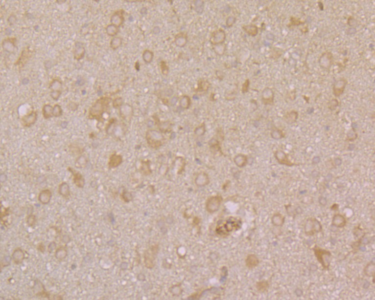
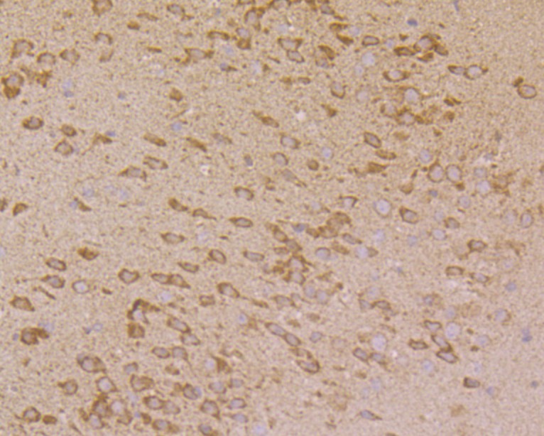
Fig5: Immunohistochemical analysis of paraffin-embedded rat brain tissue using anti-PHF8 antibody. Counter stained with hematoxylin.
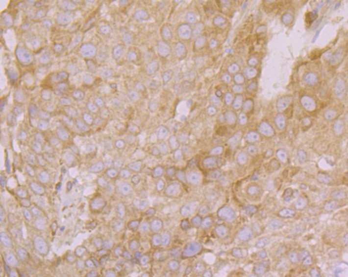
Fig6: Immunohistochemical analysis of paraffin-embedded human lung cancer tissue using anti-PHF8 antibody. Counter stained with hematoxylin.
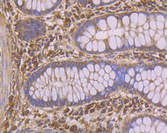
Fig7: Immunohistochemical analysis of paraffin-embedded human colon tissue using anti-PHF8 antibody. Counter stained with hematoxylin.
Fig8: Immunohistochemical analysis of paraffin-embedded mouse brain tissue using anti-PHF8 antibody. Counter stained with hematoxylin.
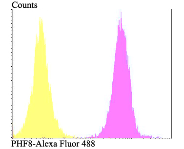
Fig9: Flow cytometric analysis of SiHa cells with PHF8 antibody at 1/100 dilution (fuchsia) compared with an unlabelled control (cells without incubation with primary antibody; yellow). Alexa Fluor 488-conjugated goat anti-rabbit IgG was used as the secon
- Background
-
References
- Qiu J et al. The X-linked mental retardation gene PHF8 is a histone demethylase involved in neuronal differentiation. Cell Res 20:908-918 (2010).
- Zhu Z et al. PHF8 is a histone H3K9me2 demethylase regulating rRNA synthesis. Cell Res 20:794-801 (2010).
Related Products / Services
Please note: All products are "FOR RESEARCH USE ONLY AND ARE NOT INTENDED FOR DIAGNOSTIC OR THERAPEUTIC USE"