
-
Product Name
Anti-PBK/SPK Mouse antibody
- Documents
-
Description
PBK/SPK Mouse monoclonal antibody
-
Tested applications
WB, IHC-P, ICC/IF, FC
-
Species reactivity
Human
-
Isotype
Mouse IgG2b
-
Preparation
Antigen: Purified recombinant fragment of human PBK expressed in E. Coli.
-
Clonality
Monoclonal
-
Formulation
Ascitic fluid containing 0.03% sodium azide.
-
Storage instructions
Store at 4°C short term. Store at -20°C long term. Avoid freeze / thaw cycle.
-
Applications
WB: 1/500 - 1/2000
IHC: 1/200 - 1/1000
ICC: 1/200 - 1/1000
FC: 1/200 - 1/400
ELISA: 1/10000
-
Validations
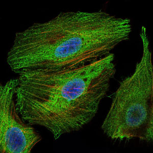
Immunofluorescence analysis of Hela cells using PBK mouse mAb (green). Blue: DRAQ5 fluorescent DNA dye. Red: Actin filaments have been labeled with Alexa Fluor-555 phalloidin.
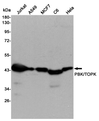
Western blot detection of PBK/TOPK in Jurkat,A549,MCF7,C6 and Hela cell lysates using PBK/TOPK mouse mAb (1:5000 diluted).Predicted band size:36KDa.Observed band size:40KDa.
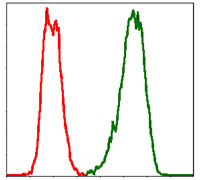
Flow cytometric analysis of Hela cells using PBK mouse mAb (green) and negative control (red).
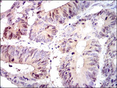
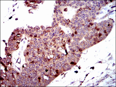
Immunohistochemical analysis of paraffin-embedded colon cancer tissues using PBK mouse mAb with DAB staining.
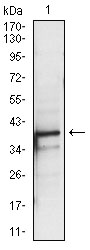
Western blot analysis using PBK mouse mAb against A431 (1) cell lysate.
Immunohistochemical analysis of paraffin-embedded ovarian cancer tissues using PBK mouse mAb with DAB staining.
-
Background
Swiss-Prot Acc.Q96KB5.This genes encodes a serine/threonine kinase related to the dual specific mitogen-activated protein kinase kinase (MAPKK) family. Evidence suggests that mitotic phosphorylation is required for its catalytic activity. This mitotic kinase may be involved in the activation of lymphoid cells and support testicular functions, with a suggested role in the process of spermatogenesis.
Related Products / Services
Please note: All products are "FOR RESEARCH USE ONLY AND ARE NOT INTENDED FOR DIAGNOSTIC OR THERAPEUTIC USE"