
-
Product Name
Anti-PAK3 antibody
- Documents
-
Description
Rabbit monoclonal antibody to PAK3
-
Tested applications
WB, ICC/IF, IHC-P, IP, FC
-
Species reactivity
Human, Mouse, Rat
-
Alternative names
ARA antibody; bPAK antibody; MRX30 antibody; MRX47 antibody; OPHN3 antibody; PAK-3 antibody; XLID30 antibody; PAK3beta antibody; beta-PAK antibody
-
Isotype
Rabbit IgG
-
Preparation
This antigen of this antibody was synthetic phospho-peptide corresponding to residues surrounding ser144 of human pak1.
-
Clonality
Monoclonal
-
Formulation
Liquid, 1*TBS (pH7.4), 0.05% BSA, 40% Glycerol. Preservative: 0.05% Sodium Azide.
-
Storage instructions
Store at +4℃ after thawing. Aliquot store at -20℃ or -80℃. Avoid repeated freeze / thaw cycles.
-
Applications
WB: 1:1,000-1:2,000
ICC/IF: 1:50-1:200
IHC-P: 1:50-1:200
FC: 1:50-1:100
IP: Use at an assay dependent concentration.
-
Validations
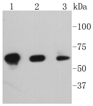
Fig1:; Western blot analysis of Phospho-PAK1(S144)+PAK2(S141)+PAK3(S139) on different lysates. Proteins were transferred to a PVDF membrane and blocked with 5% BSA in PBS for 1 hour at room temperature. The primary antibody ( 1/500) was used in 5% BSA at room temperature for 2 hours. Goat Anti-Rabbit IgG - HRP Secondary Antibody (HA1001) at 1:5,000 dilution was used for 1 hour at room temperature.; Positive control:; Lane 1: Hela cell lysate; Lane 2: NIH/3T3 cell lysate; Lane 2: SH-SY5Y cell lysate
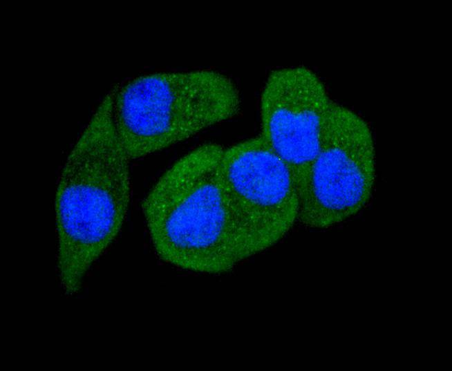
Fig2:; ICC staining of Phospho-PAK1(S144)+PAK2(S141)+PAK3(S139) in Hela cells (green). Formalin fixed cells were permeabilized with 0.1% Triton X-100 in TBS for 10 minutes at room temperature and blocked with 1% Blocker BSA for 15 minutes at room temperature. Cells were probed with the primary antibody ( 1/50) for 1 hour at room temperature, washed with PBS. Alexa Fluor®488 Goat anti-Rabbit IgG was used as the secondary antibody at 1/1,000 dilution. The nuclear counter stain is DAPI (blue).
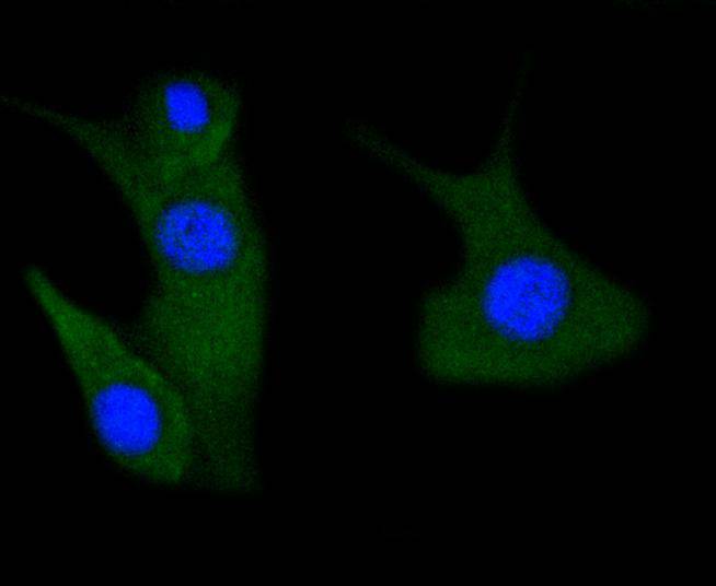
Fig3:; ICC staining of Phospho-PAK1(S144)+PAK2(S141)+PAK3(S139) in NIH/3T3 cells (green). Formalin fixed cells were permeabilized with 0.1% Triton X-100 in TBS for 10 minutes at room temperature and blocked with 1% Blocker BSA for 15 minutes at room temperature. Cells were probed with the primary antibody ( 1/50) for 1 hour at room temperature, washed with PBS. Alexa Fluor®488 Goat anti-Rabbit IgG was used as the secondary antibody at 1/1,000 dilution. The nuclear counter stain is DAPI (blue).
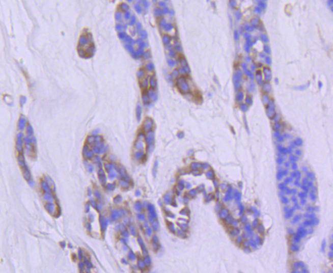
Fig4:; Immunohistochemical analysis of paraffin-embedded human liver carcinoma tissue using anti-Phospho-PAK1(S144)+PAK2(S141)+PAK3(S139) antibody. The section was pre-treated using heat mediated antigen retrieval with Tris-EDTA buffer (pH 8.0-8.4) for 20 minutes.The tissues were blocked in 5% BSA for 30 minutes at room temperature, washed with ddH; 2; O and PBS, and then probed with the primary antibody ( 1/50) for 30 minutes at room temperature. The detection was performed using an HRP conjugated compact polymer system. DAB was used as the chromogen. Tissues were counterstained with hematoxylin and mounted with DPX.
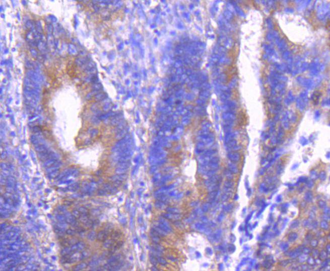
Fig5:; Immunohistochemical analysis of paraffin-embedded human colon carcinoma tissue using anti-Phospho-PAK1(S144)+PAK2(S141)+PAK3(S139) antibody. The section was pre-treated using heat mediated antigen retrieval with Tris-EDTA buffer (pH 8.0-8.4) for 20 minutes.The tissues were blocked in 5% BSA for 30 minutes at room temperature, washed with ddH; 2; O and PBS, and then probed with the primary antibody ( 1/50) for 30 minutes at room temperature. The detection was performed using an HRP conjugated compact polymer system. DAB was used as the chromogen. Tissues were counterstained with hematoxylin and mounted with DPX.
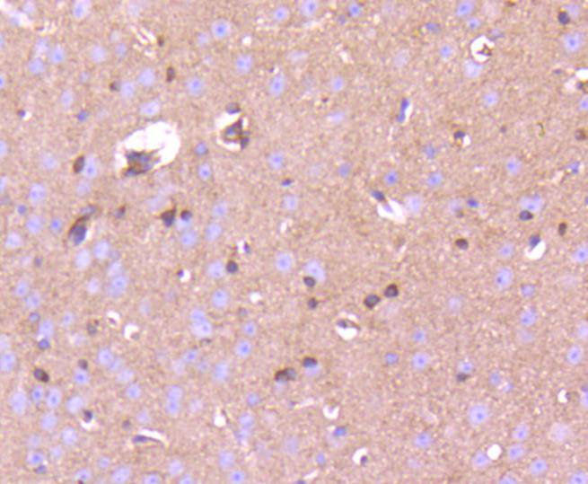
Fig6:; Immunohistochemical analysis of paraffin-embedded mouse brain tissue using anti-Phospho-PAK1(S144)+PAK2(S141)+PAK3(S139) antibody. The section was pre-treated using heat mediated antigen retrieval with Tris-EDTA buffer (pH 8.0-8.4) for 20 minutes.The tissues were blocked in 5% BSA for 30 minutes at room temperature, washed with ddH; 2; O and PBS, and then probed with the primary antibody ( 1/50) for 30 minutes at room temperature. The detection was performed using an HRP conjugated compact polymer system. DAB was used as the chromogen. Tissues were counterstained with hematoxylin and mounted with DPX.
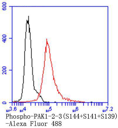
Fig7:; Flow cytometric analysis of Phospho-PAK1(S144)+PAK2(S141)+PAK3(S139) was done on NIH/3T3 cells. The cells were fixed, permeabilized and stained with the primary antibody ( 1/50) (red). After incubation of the primary antibody at room temperature for an hour, the cells were stained with a Alexa Fluor 488-conjugated Goat anti-Rabbit IgG Secondary antibody at 1/1000 dilution for 30 minutes.Unlabelled sample was used as a control (cells without incubation with primary antibody; black).
- Background
-
References
- El-Baba C et al. Thymoquinone-induced conformational changes of PAK1 interrupt prosurvival MEK-ERK signaling in colorectal cancer. Mol Cancer 13:201 (2014).
- Freeman MC et al. Coronaviruses induce entry-independent, continuous macropinocytosis. MBio 5:e01340-14 (2014).
Related Products / Services
Please note: All products are "FOR RESEARCH USE ONLY AND ARE NOT INTENDED FOR DIAGNOSTIC OR THERAPEUTIC USE"