
-
Product Name
Anti-NELFA antibody
- Documents
-
Description
Mouse monoclonal antibody to NELFA
-
Tested applications
WB, ICC, IHC-P, FC
-
Species reactivity
Human, Rat
-
Alternative names
WHSC2 antibody; NELF-A antibody; P/OKcl.15 antibody
-
Isotype
Mouse IgG2b
-
Preparation
This antigen of this antibody was recombinant protein
-
Clonality
Monoclonal
-
Formulation
Liquid, 1*TBS (pH7.4), 1%BSA, 40%Glycerol. Preservative: 0.05% Sodium Azide.
-
Storage instructions
Store at +4℃ after thawing. Aliquot store at -20℃ or -80℃. Avoid repeated freeze / thaw cycles.
-
Applications
WB: 1:500-1:2,000
ICC: 1:50-1:200
IHC-P: 1:100-1:200
FC: 1:100-1:200
-
Validations
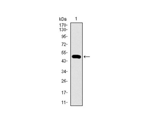
Fig1: Western blot analysis of WHSC2 on human WHSC2 recombinant protein using anti-WHSC2 antibody at 1/1,000 dilution.
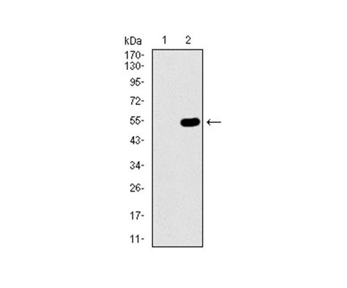
Fig2: Western blot analysis of WHSC2 on HEK293 (1) and WHSC2-hIgGFc transfected HEK293 (2) cell lysate using anti-WHSC2 antibody at 1/1,000 dilution.
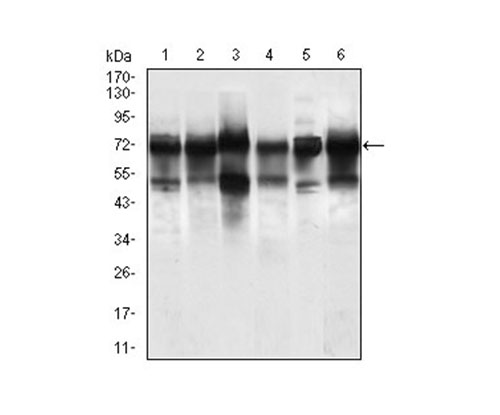
Fig3: Western blot analysis of WHSC2 on different cell lysate using anti-WHSC2 antibody at 1/1,000 dilution.; Positive control:; Line1:Jurkat Line2:HeLa Line3:HEK293; Line4:Rat brain Line5:A549 Line6:SPC-A-1
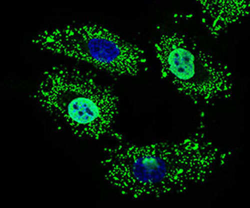
Fig4: ICC staining WHSC2 (green) in HeLa cells. The nuclear counter stain is DAPI (blue). Cells were fixed in paraformaldehyde, permeabilised with 0.25% Triton X100/PBS.
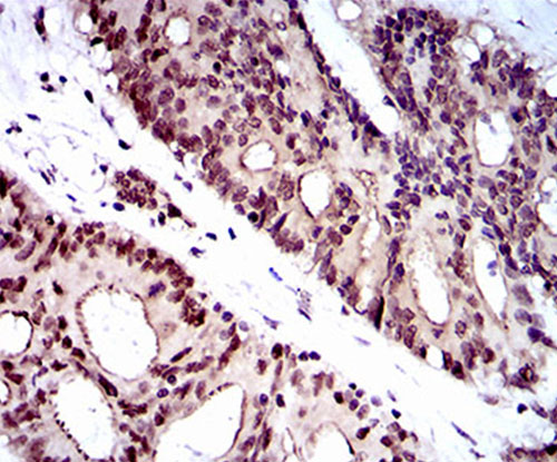
Fig5: Immunohistochemical analysis of paraffin-embedded human colon cancer tissue using anti-WHSC2 antibody. Counter stained with hematoxylin.
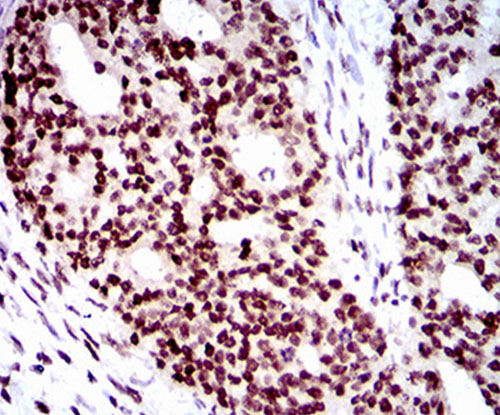
Fig6: Immunohistochemical analysis of paraffin-embedded human ovarian cancer tissue using anti-WHSC2 antibody. Counter stained with hematoxylin.
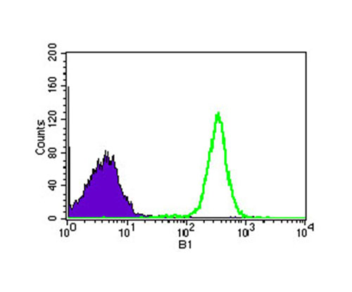
Fig7: Flow cytometric analysis of HEK293 cells with WHSC2 antibody at 1/100 dilution (green) compared with an unlabelled control (cells without incubation with primary antibody; purple).
- Background
-
References
- Kerzendorfer C et al. Characterizing the functional consequences of haploinsufficiency of NELF-A (WHSC2) and SLBP identifies novel cellular phenotypes in Wolf-Hirschhorn syndrome. Hum Mol Genet 21(10):2181-93 (2012).
- Yung TM et al. Cellular dynamics of the negative transcription elongation factor NELF. Exp Cell Res 315(10):1693-705 (2009).
Related Products / Services
Please note: All products are "FOR RESEARCH USE ONLY AND ARE NOT INTENDED FOR DIAGNOSTIC OR THERAPEUTIC USE"