
-
Product Name
Anti-LRP3 antibody
- Documents
-
Description
Mouse monoclonal antibody to LRP3
-
Tested applications
WB, ICC, FC
-
Species reactivity
Human
-
Isotype
Mouse IgG1
-
Preparation
This antigen of this antibody was purified recombinant fragment of human lrp3 (aa: extra 43-184) expressed in e. coli.
-
Clonality
Monoclonal
-
Formulation
Liquid, 1*PBS with 0.05% sodium azide.
-
Storage instructions
Store at +4℃ after thawing. Aliquot store at -20℃. Avoid repeated freeze / thaw cycles.
-
Applications
WB: 1:500-1:2,000
ICC: 1:50-1:200
FC: 1:100-1:200
-
Validations
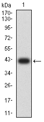
Fig1: Western blot analysis of LRP3 against human LRP3 (AA: extra 43-184) recombinant protein. Proteins were transferred to a PVDF membrane and blocked with 5% BSA in PBS for 1 hour at room temperature. The primary antibody ( 1/500) was used in 5% BSA at room temperature for 2 hours. Goat Anti-Mouse IgG - HRP Secondary Antibody at 1:5,000 dilution was used for 1 hour at room temperature.
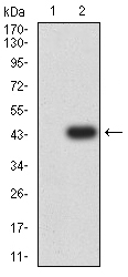
Fig2: Western blot analysis of 175076# against HEK293 (1) and LRP3 (AA: extra 43-184)-hIgGFc transfected HEK293 (2) cell lysate.Proteins were transferred to a PVDF membrane and blocked with 5% BSA in PBS for 1 hour at room temperature. The primary antibody ( 1/500) was used in 5% BSA at room temperature for 2 hours. Goat Anti-Mouse IgG - HRP Secondary Antibody at 1:5,000 dilution was used for 1 hour at room temperature.
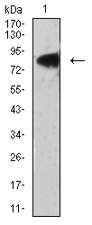
Fig3: Western blot analysis of 175076# against PANC-1 (1) cell lysate.Proteins were transferred to a PVDF membrane and blocked with 5% BSA in PBS for 1 hour at room temperature. The primary antibody ( 1/500) was used in 5% BSA at room temperature for 2 hours. Goat Anti-Mouse IgG - HRP Secondary Antibody at 1:5,000 dilution was used for 1 hour at room temperature.
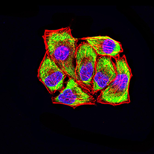
Fig4: Immunocytochemistry staining of LRP3 in Hela cells (green). Formalin fixed cells were permeabilized with 0.1% Triton X-100 in TBS for 10 minutes at room temperature and blocked with 1% Blocker BSA for 15 minutes at room temperature. Cells were probed with the primary antibody ( 1/100) for 1 hour at room temperature, washed with PBS. Alexa Fluor®488 Goat anti-Mouse IgG was used as the secondary antibody at 1/1,000 dilution. The nuclear counter stain is DAPI (blue), Actin filaments have been labeled with Alexa Fluor- 555 phalloidin (red).
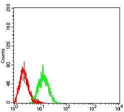
Fig5: Flow cytometric analysis of LRP3 was done on HL-60 cells. The cells were fixed, permeabilized and stained with the primary antibody ( 1/100) (green). After incubation of the primary antibody at room temperature for an hour, the cells were stained with a Alexa Fluor 488-conjugated goat anti-Mouse IgG Secondary antibody at 1/500 dilution for 30 minutes. Unlabelled sample was used as a control (cells without incubation with primary antibody; red).
- Background
-
References
- Stem Cell Res. 2017 Apr;20:94-104.
Related Products / Services
Please note: All products are "FOR RESEARCH USE ONLY AND ARE NOT INTENDED FOR DIAGNOSTIC OR THERAPEUTIC USE"