
-
Product Name
Anti-IL31 antibody
- Documents
-
Description
Mouse monoclonal antibody to IL31
-
Tested applications
WB, IHC-P, ICC
-
Species reactivity
Human
-
Alternative names
IL-31 antibody
-
Isotype
Mouse IgG3
-
Preparation
This antigen of this antibody was recombinant full length protein corresponding to human il18.
-
Clonality
Monoclonal
-
Formulation
Liquid, 1*PBS (pH7.4), 0.2% BSA, 50% Glycerol. Preservative: 0.05% Sodium Azide.
-
Storage instructions
Store at +4℃ after thawing. Aliquot store at -20℃. Avoid repeated freeze / thaw cycles.
-
Applications
WB: 1:500
ICC: 1:100
IHC-P: 1:50-1:200
-
Validations
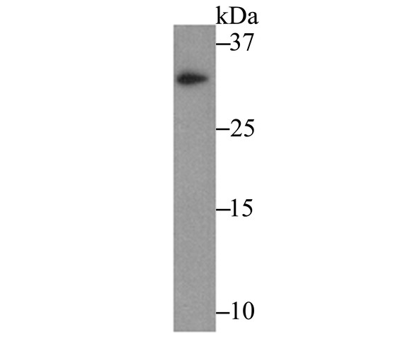
Fig1: Western blot analysis of IL-31 on recombinant protein lysate using anti-IL-31 antibody at 1/500 dilution.
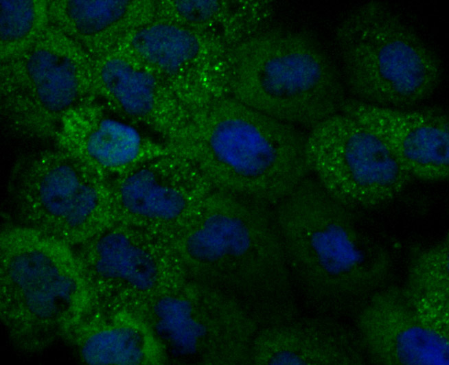
Fig2: ICC staining IL-31 (green) in A431 cells. The nuclear counter stain is DAPI (blue). Cells were fixed in paraformaldehyde, permeabilised with 0.25% Triton X100/PBS.
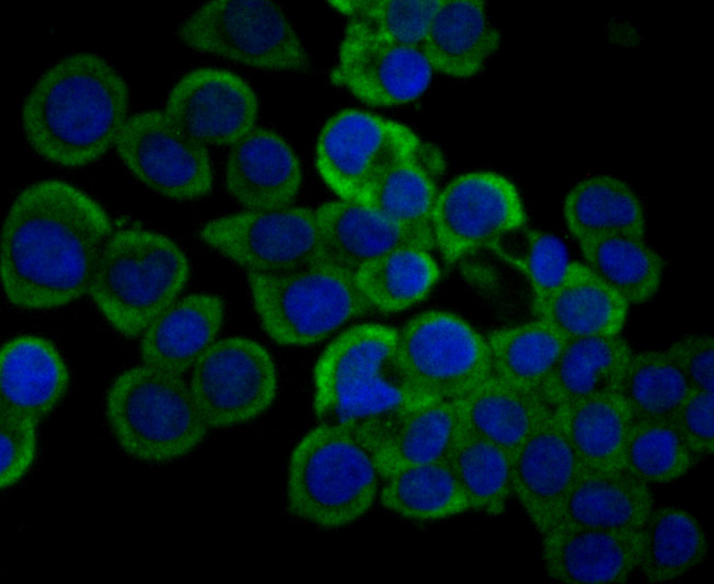
Fig3: ICC staining IL-31 (green) in LOVO cells. The nuclear counter stain is DAPI (blue). Cells were fixed in paraformaldehyde, permeabilised with 0.25% Triton X100/PBS.
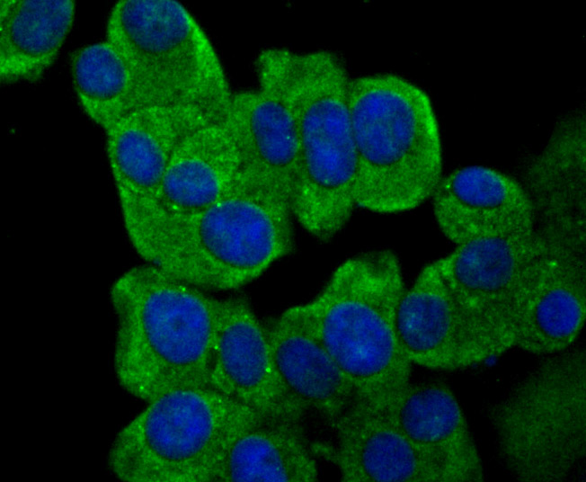
Fig4: ICC staining IL-31 (green) in MCF-7 cells. The nuclear counter stain is DAPI (blue). Cells were fixed in paraformaldehyde, permeabilised with 0.25% Triton X100/PBS.
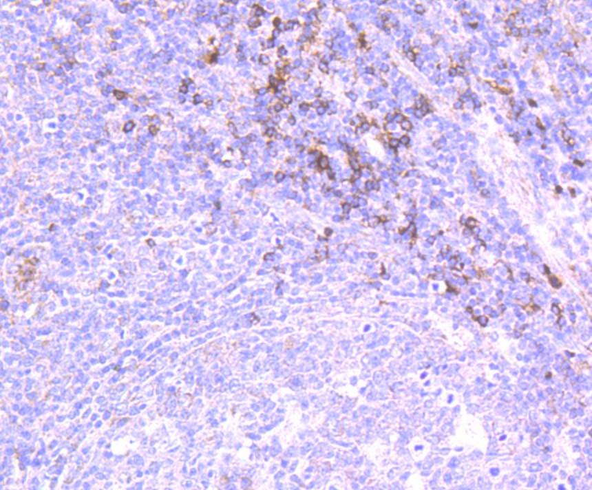
Fig5: Immunohistochemical analysis of paraffin-embedded human tonsil tissue using anti-IL-31 antibody. Counter stained with hematoxylin.
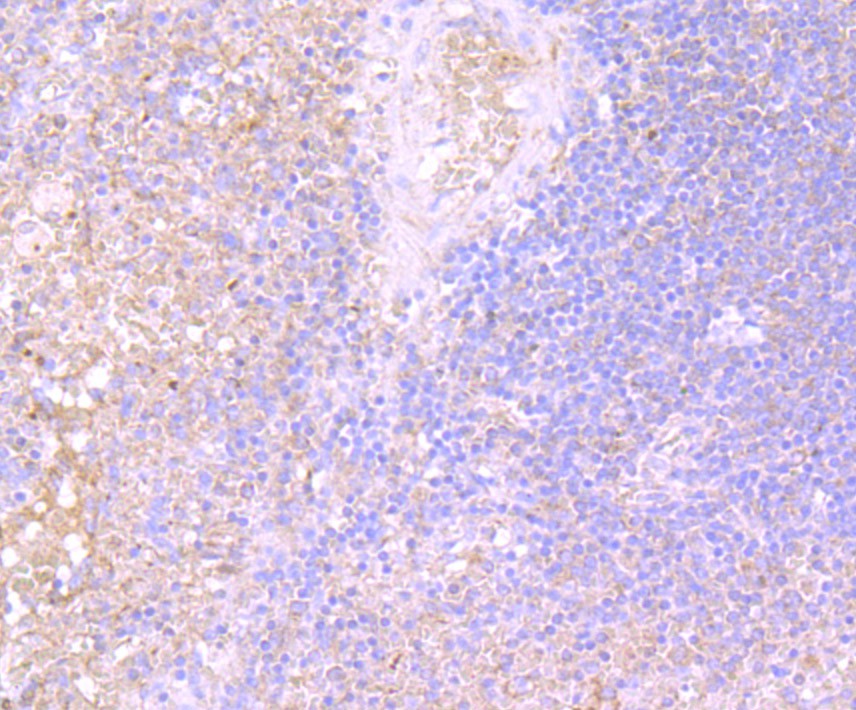
Fig6: Immunohistochemical analysis of paraffin-embedded human spleen tissue using anti-IL-31 antibody. Counter stained with hematoxylin.
- Background
-
References
- Dillon S R et al. Interleukin 31, a cytokine produced by activated T cells, induces dermatitis in mice. Nat Immunol 5:752-760 (2004).
- Neis M M et al. Enhanced expression levels of IL-31 correlate with IL-4 and IL-13 in atopic and allergic contact dermatitis. J Allergy Clin Immunol 118:930-937(2006).
Related Products / Services
Please note: All products are "FOR RESEARCH USE ONLY AND ARE NOT INTENDED FOR DIAGNOSTIC OR THERAPEUTIC USE"