
-
Product Name
Anti-Il12a antibody
- Documents
-
Description
Rabbit polyclonal antibody to Il12a
-
Tested applications
WB, IHC-P, FC
-
Species reactivity
Mouse, Rat
-
Alternative names
p3 antibody; p35 antibody; IL-12 antibody; Ll12a antibody; Il-12a antibody; IL-12p35 antibody
-
Isotype
Rabbit IgG
-
Preparation
This antigen of this antibody was klh conjugated synthetic peptide derived from mouse il-12 51-150/215
-
Clonality
Polyclonal
-
Formulation
Liquid, 0.01M TBS(pH7.4) with 1% BSA, 0.03% Proclin300 and 50% Glycerol.
-
Storage instructions
Store at -20℃ for one year. Avoid repeated freeze/thaw cycles. The lyophilized antibody is stable at room temperature for at least one month and for greater than a year when kept at -20℃. When reconstituted in sterile pH 7.4 0.01M PBS or diluent of antibody the antibody is stable for at least two weeks at 2-4℃.
-
Applications
WB:1:500-2000
IHC-P:1:400-800
FC:1μg/Test
-
Validations
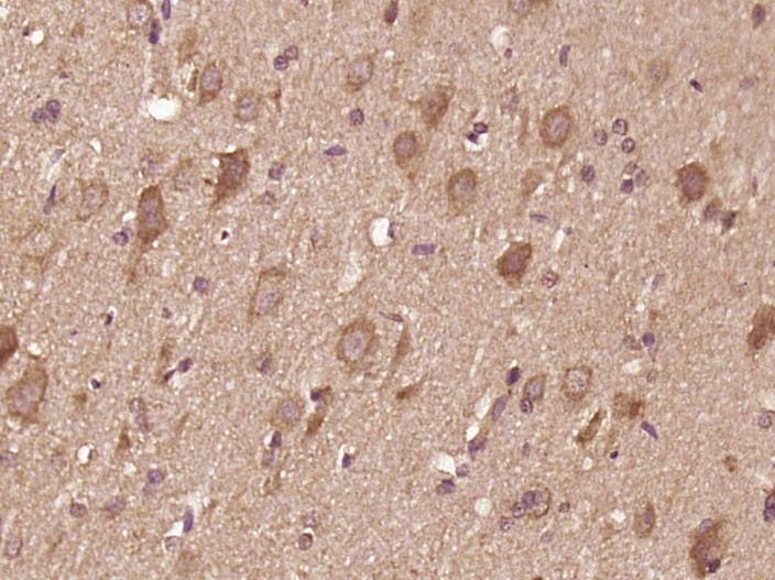
Fig1: Paraformaldehyde-fixed, paraffin embedded (rat brain tissue); Antigen retrieval by boiling in sodium citrate buffer (pH6.0) for 15min; Block endogenous peroxidase by 3% hydrogen peroxide for 20 minutes; Blocking buffer (normal goat serum) at 37℃ for 30min; Antibody incubation with (IL12) Polyclonal Antibody, Unconjugated at 1:400 overnight at 4℃, followed by operating according to SP Kit(Rabbit) (sp-0023) instructionsand DAB staining.
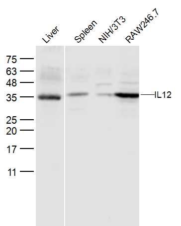
Fig2: Sample:; Liver (Mouse) Lysate at 40 ug; Spleen (Mouse) Lysate at 40 ug; NIH/3T3 (Mouse) CellLysate at 30 ug; RAW246.7 (Mouse) CellLysate at 30 ug; Primary: Anti- IL12 at 1/300 dilution; Secondary: IRDye800CW Goat Anti-Rabbit IgG at 1/20000 dilution; Predicted band size: 22 kD; Observed band size: 35/36 kD
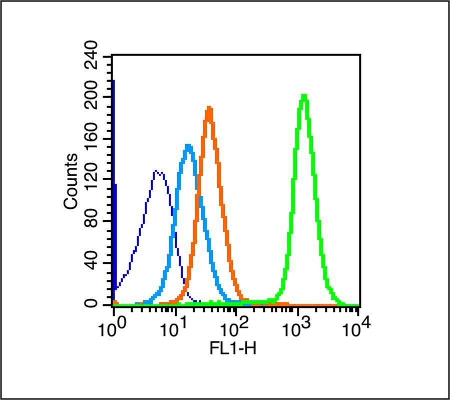
Fig3: Blank control (blue line): Mouse spleen (blue).; Primary Antibody (green line): Rabbit Anti- IL12 antibody ; Dilution: 1μg /10^6 cells;; Isotype Control Antibody (orange line): Rabbit IgG .; Secondary Antibody (white blue line): Goat anti-rabbit IgG-FITC; Dilution: 1μg /test.; Protocol; The cells were fixed with 70% ice-cold methanol overnight at 4℃. Cells stained with Primary Antibody for 30 min at room temperature. The cells were then incubated in 1 X PBS/2%BSA/10% goat serum to block non-specific protein-protein interactions followed by the antibody for 15 min at room temperature. The secondary antibody used for 40 min at room temperature. Acquisition of 20,000 events was performed.
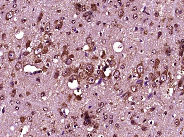
Fig4: Paraformaldehyde-fixed, paraffin embedded (Mouse brain); Antigen retrieval by boiling in sodium citrate buffer (pH6.0) for 15min; Block endogenous peroxidase by 3% hydrogen peroxide for 20 minutes; Blocking buffer (normal goat serum) at 37℃ for 30min; Antibody incubation with (IL12) Polyclonal Antibody, Unconjugated at 1:400 overnight at 4℃, followed by operating according to SP Kit(Rabbit) (sp-0023) instructions and DAB staining.
- Background
Related Products / Services
Please note: All products are "FOR RESEARCH USE ONLY AND ARE NOT INTENDED FOR DIAGNOSTIC OR THERAPEUTIC USE"