
-
Product Name
Anti-Hnf1a antibody
- Documents
-
Description
Rabbit polyclonal antibody to Hnf1a
-
Tested applications
WB, IHC-P, FC
-
Species reactivity
Human, Mouse, Rat, Chicken, Dog, Pig, Cow, Zebrafish, Sheep
-
Alternative names
HNF1 antibody; Lfb1 antibody; Tcf1 antibody; LF-B1 antibody
-
Isotype
Rabbit IgG
-
Preparation
This antigen of this antibody was klh conjugated synthetic peptide derived from human hnf1 201-350/628
-
Clonality
Polyclonal
-
Formulation
Liquid, 0.01M TBS(pH7.4) with 1% BSA, 0.03% Proclin300 and 50% Glycerol.
-
Storage instructions
Store at -20℃ for one year. Avoid repeated freeze/thaw cycles. The lyophilized antibody is stable at room temperature for at least one month and for greater than a year when kept at -20℃. When reconstituted in sterile pH 7.4 0.01M PBS or diluent of antibody the antibody is stable for at least two weeks at 2-4℃.
-
Applications
WB:1:500-2000
IHC-P:1:400-800
FC:3ug/test
-
Validations
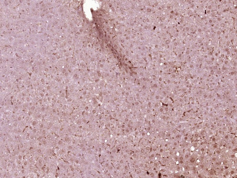
Fig1: Paraformaldehyde-fixed, paraffin embedded (Mouse liver); Antigen retrieval by boiling in sodium citrate buffer (pH6.0) for 15min; Block endogenous peroxidase by 3% hydrogen peroxide for 20 minutes; Blocking buffer (normal goat serum) at 37℃ for 30min; Antibody incubation with (HNF1A) Polyclonal Antibody, Unconjugated at 1:400 overnight at 4℃, followed by operating according to SP Kit(Rabbit) (sp-0023) instructionsand DAB staining.
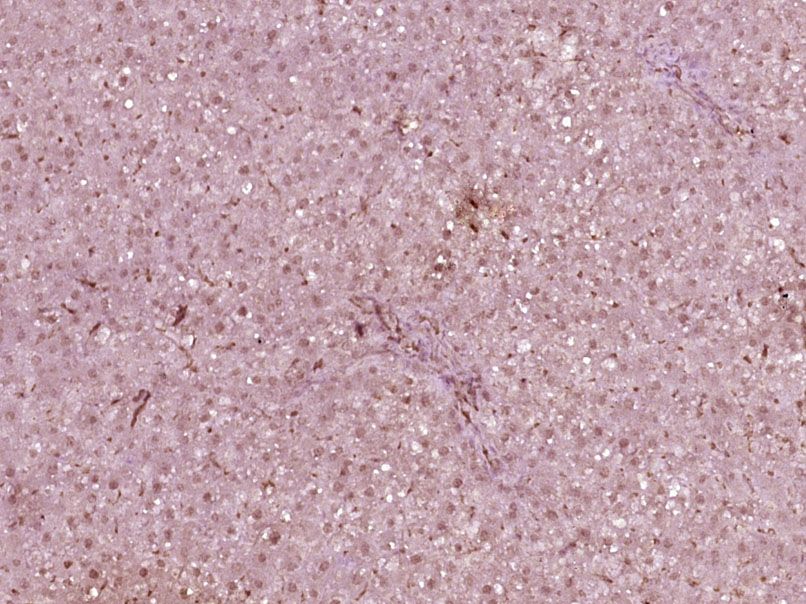
Fig2: Paraformaldehyde-fixed, paraffin embedded (Rat liver); Antigen retrieval by boiling in sodium citrate buffer (pH6.0) for 15min; Block endogenous peroxidase by 3% hydrogen peroxide for 20 minutes; Blocking buffer (normal goat serum) at 37℃ for 30min; Antibody incubation with (HNF1A) Polyclonal Antibody, Unconjugated at 1:400 overnight at 4℃, followed by operating according to SP Kit(Rabbit) (sp-0023) instructionsand DAB staining.
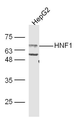
Fig3: Sample:; HepG2 Cell (Human) Lysate at 30 ug; Primary: Anti-HNF1 (Bs-1405R) at 1/300 dilution; Secondary: IRDye800CW Goat Anti-Rabbit IgG at 1/20000 dilution; Predicted band size: 67 kD; Observed band size: 67 kD
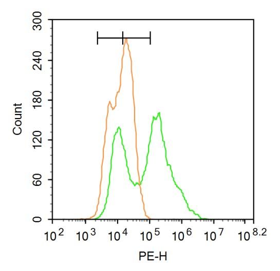
Fig4: Blank control:A549.; Primary Antibody (green line): Rabbit Anti-HNF1A antibody ; Dilution: 1μg /10^6 cells;; Isotype Control Antibody (orange line): Rabbit IgG .; Secondary Antibody : Goat anti-rabbit IgG-PE; Dilution: 3μg /test.; Protocol; The cells were fixed with 4% PFA (10min at room temperature)and then permeabilized with 90% ice-cold methanol for 20 min at-20℃. The cells were then incubated in 5% BSA to block non-specific protein-protein interactions for 30 min at at room temperature .Cells stained with Primary Antibody for 30 min at room temperature. The secondary antibody used for 40 min at room temperature. Acquisition of 20,000 events was performed.
- Background
Related Products / Services
Please note: All products are "FOR RESEARCH USE ONLY AND ARE NOT INTENDED FOR DIAGNOSTIC OR THERAPEUTIC USE"