

-
Product Name
Anti-GPX1 (C-term) Rabbit antibody
- Documents
-
Description
GPX1 (C-term) Rabbit polyclonal antibody
-
Tested applications
FC, WB, IHC-P, ICC/IF
-
Species reactivity
Human, Mouse, Rat
-
Isotype
Rabbit IgG
-
Preparation
Antigen: This GPX1 antibody is generated from rabbits immunized with a KLH conjugated synthetic peptide between 164-193 amino acids from the C-terminal region of human GPX1.
-
Clonality
Polyclonal
-
Formulation
Purified polyclonal antibody supplied in PBS with 0.09% (W/V) sodium azide. This antibody is purified through a protein A column, followed by peptide affinity purification.
-
Applications
WB::1:2000ICC::1:10~50IHC::1:10~50
-
Validations
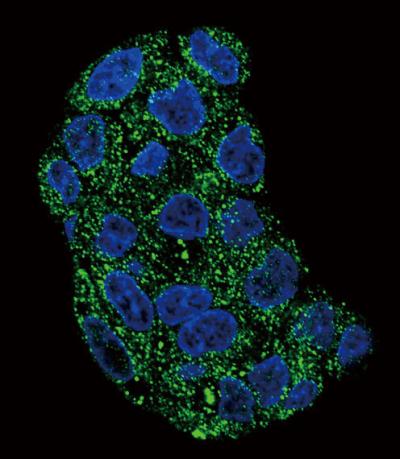
Confocal immunofluorescent analysis of GPX1 Antibody (C-term)(Cat#169114) with HepG2 cell followed by Alexa Fluor 488-conjugated goat anti-rabbit lgG (green). DAPI was used to stain the cell nuclear (blue).
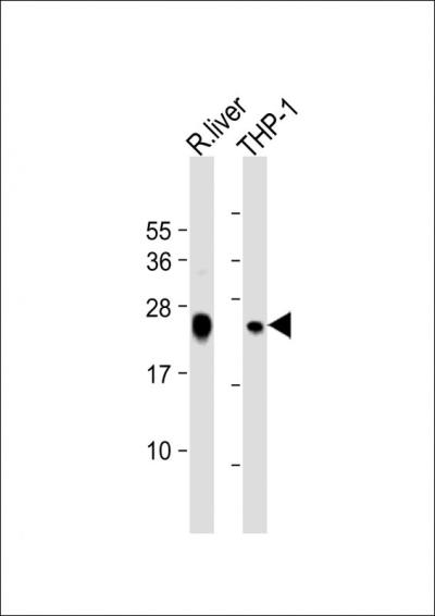
All lanes: Anti-GPX1 Antibody (C-term) at 1:2000 dilutionnLane 1: rat liver lysatenLane 2: THP-1 whole cell lysatennLysates/proteins at 20 u03bcg per lane. nnSecondarynGoat Anti-Rabbit IgG, (H+L), Peroxidase conjugated at 1/10000 dilution. nnPredicted band size: 22 kDannBlocking/Dilution buffer: 5% NFDM/TBST.
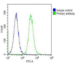
Overlay histogram showing Hela cells stained with 169114 (green line). The cells were fixed with 2% paraformaldehyde (10 min) and then permeabilized with 90% methanol for 10 min. The cells were then icubated in 2% bovine serum albumin to block non-spec
Western blot analysis of lysates from THP-1 cell line uff0cmouse liver and rat liver tissue (from left to right),using GPX1 Antibody (C-term)(Cat. #169114).169114 was diluted at 1:1000 at each lane. A goat anti-rabbit IgG H&L(HRP) at 1:5000 dilution was used as the secondary antibody.Lysates at 35ug per lane.
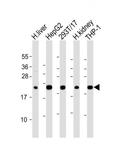
All lanes: Anti-GPX1 Antibody (C-term) at 1:2000 dilutionnLane 1: human liver lysatenLane 2: HepG2 whole cell lysatenLane 3: 293T/17 whole cell lysatenLane 4: human kidney lysatenLane 5: THP-1 whole cell lysatennLysates/proteins at 20 u03bcg per lane. nnSecondarynGoat Anti-Rabbit IgG, (H+L), Peroxidase conjugated at 1/10000 dilution. nnPredicted band size: 22 kDannBlocking/Dilution buffer: 5% NFDM/TBST.
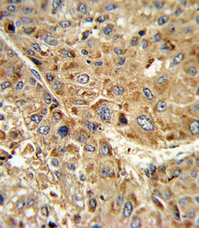
Formalin-fixed and paraffin-embedded human hepatocarcinoma reacted with GPX1 Antibody (C-term), which was peroxidase-conjugated to the secondary antibody, followed by DAB staining. This data demonstrates the use of this antibody for immunohistochemistry;
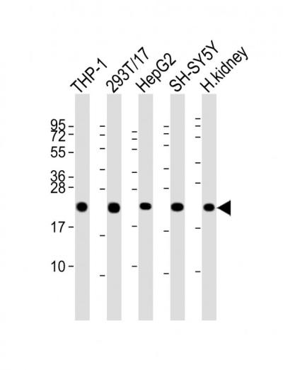
All lanes: Anti-GPX1 Antibody (C-term) at 1:2000 dilutionnLane 1: THP-1 whole cell lysatenLane 2: 293T/17 whole cell lysatenLane 3: HepG2 whole cell lysatenLane 4: SH-SY5Y whole cell lysatenLane 5: human kidney lysatennLysates/proteins at 20 u03bcg per lane. nnSecondarynGoat Anti-Rabbit IgG, (H+L), Peroxidase conjugated at 1/10000 dilution. nnPredicted band size: 22 kDannBlocking/Dilution buffer: 5% NFDM/TBST.
-
Background
Swiss-Prot Acc.P07203.
Related Products / Services
Please note: All products are "FOR RESEARCH USE ONLY AND ARE NOT INTENDED FOR DIAGNOSTIC OR THERAPEUTIC USE"