
-
Product Name
Anti-GH1 antibody
- Documents
-
Description
Rabbit monoclonal antibody to GH1
-
Tested applications
WB, IP, IHC-P
-
Species reactivity
Human
-
Alternative names
GH antibody; GHN antibody; GH-N antibody; GHB5 antibody; IGHD2 antibody; hGH-N antibody; IGHD1A antibody; IGHD1B antibody
-
Isotype
Rabbit IgG
-
Preparation
This antigen of this antibody was synthetic peptide within human growth hormone aa 72-106 / 217.
-
Clonality
Monoclonal
-
Formulation
Liquid, 1*TBS (pH7.4), 0.05% BSA, 40% Glycerol. Preservative: 0.05% Sodium Azide.
-
Storage instructions
Store at +4℃ after thawing. Aliquot store at -20℃ or -80℃. Avoid repeated freeze / thaw cycles.
-
Applications
WB: 1:1,000-1:5,000
IHC-P: 1:50-1:200
-
Validations
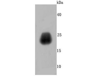
Fig1:; Western blot analysis of Growth Hormone on human placenta tissue lysates. Proteins were transferred to a PVDF membrane and blocked with 5% BSA in PBS for 1 hour at room temperature. The primary antibody ( 1/500) was used in 5% BSA at room temperature for 2 hours. Goat Anti-Rabbit IgG - HRP Secondary Antibody (HA1001) at 1:200,000 dilution was used for 1 hour at room temperature.
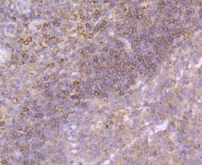
Fig2:; Immunohistochemical analysis of paraffin-embedded human tonsil tissue using anti-Growth Hormone antibody. The section was pre-treated using heat mediated antigen retrieval with Tris-EDTA buffer (pH 9.0) for 20 minutes.The tissues were blocked in 1% BSA for 30 minutes at room temperature, washed with ddH; 2; O and PBS, and then probed with the primary antibody ( 1/50) for 30 minutes at room temperature. The detection was performed using an HRP conjugated compact polymer system. DAB was used as the chromogen. Tissues were counterstained with hematoxylin and mounted with DPX.
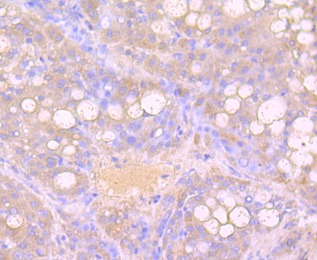
Fig3:; Immunohistochemical analysis of paraffin-embedded human liver carcinoma tissue using anti-Growth Hormone antibody. The section was pre-treated using heat mediated antigen retrieval with Tris-EDTA buffer (pH 9.0) for 20 minutes.The tissues were blocked in 1% BSA for 30 minutes at room temperature, washed with ddH; 2; O and PBS, and then probed with the primary antibody ( 1/50) for 30 minutes at room temperature. The detection was performed using an HRP conjugated compact polymer system. DAB was used as the chromogen. Tissues were counterstained with hematoxylin and mounted with DPX.
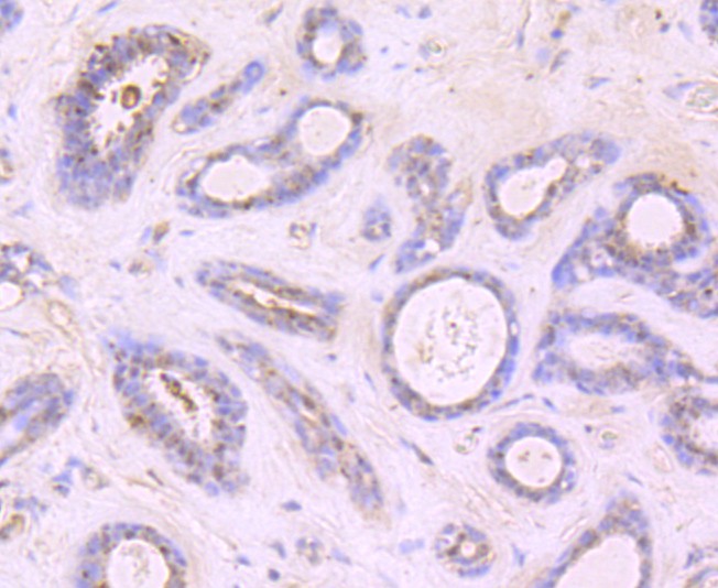
Fig4:; Immunohistochemical analysis of paraffin-embedded human breast carcinoma tissue using anti-Growth Hormone antibody. The section was pre-treated using heat mediated antigen retrieval with Tris-EDTA buffer (pH 9.0) for 20 minutes.The tissues were blocked in 1% BSA for 30 minutes at room temperature, washed with ddH; 2; O and PBS, and then probed with the primary antibody ( 1/50) for 30 minutes at room temperature. The detection was performed using an HRP conjugated compact polymer system. DAB was used as the chromogen. Tissues were counterstained with hematoxylin and mounted with DPX.
- Background
-
References
- Kang Y et al. Tim29 is a novel subunit of the human TIM22 translocase and is involved in complex assembly and stability. Elife 5:N/A (2016).
- Donti TR et al. Expanding the phenotypic spectrum of Succinyl-CoA ligase deficiency through functional validation of a new SUCLG1 variant. Mol Genet Metab 119:68-74 (2016).
Related Products / Services
Please note: All products are "FOR RESEARCH USE ONLY AND ARE NOT INTENDED FOR DIAGNOSTIC OR THERAPEUTIC USE"