
-
Product Name
Anti-Gap43 antibody
- Documents
-
Description
Rabbit polyclonal antibody to Gap43
-
Tested applications
WB, ICC, IHC-P, FC
-
Species reactivity
Human, Mouse, Rat
-
Alternative names
Ba antibody; B-50 antibody; GAP- antibody; Basp2 antibody; GAP-43 antibody
-
Isotype
Rabbit IgG
-
Preparation
This antigen of this antibody was peptide
-
Clonality
Polyclonal
-
Formulation
Liquid, 1*PBS (pH7.4), 0.2% BSA, 40% Glycerol. Preservative: 0.05% Sodium Azide.
-
Storage instructions
Store at +4℃ after thawing. Aliquot store at -20℃ or -80℃. Avoid repeated freeze / thaw cycles.
-
Applications
WB: 1:1,000
ICC: 1:200
IHC-P: 1:200
FC: 1:100-1:200
-
Validations
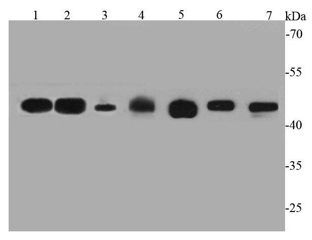
Fig1: Western blot analysis of GAP43 on different cell lysates using anti-GAP43 antibody at 1/1000 dilution.; Positive control:; Lane 1: Rat brain; Lane 2: Mouse brain; Lane 3: Mouse heart; Lane 4: Human skeletal muscle; Lane 5: N2A; Lane 6: A172; Lane 7: Human heart
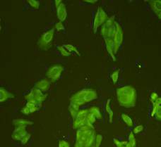
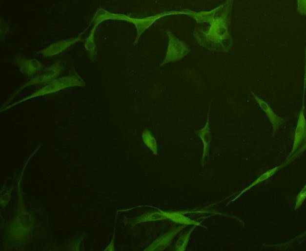
Fig2: ICC staining GAP43 in Hela cells (green). Cells were fixed in paraformaldehyde, permeabilised with 0.25% Triton X100/PBS.
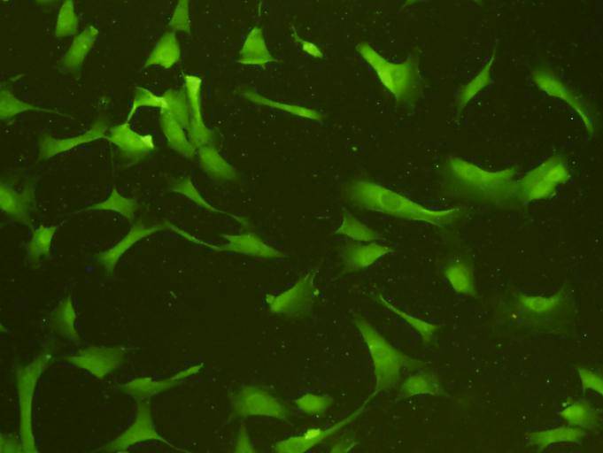
Fig3: ICC staining GAP43 in NIH/3T3 cells (green). Cells were fixed in paraformaldehyde, permeabilised with 0.25% Triton X100/PBS.
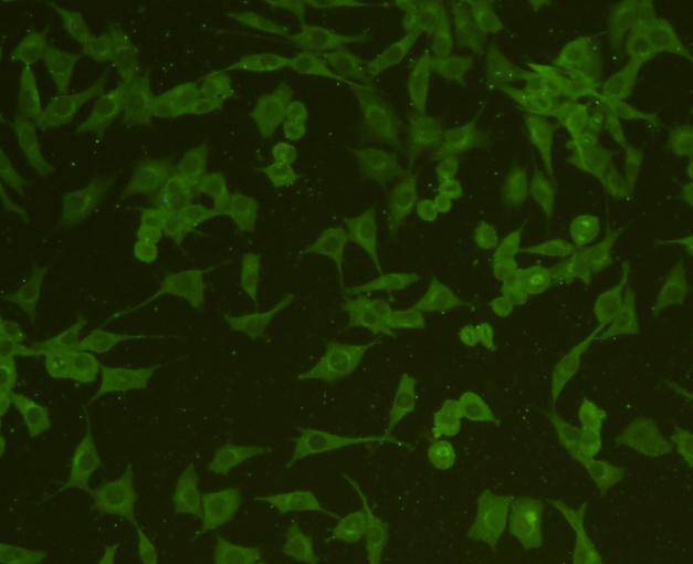
Fig4: ICC staining GAP43 in SHG-44 cells (green). Cells were fixed in paraformaldehyde, permeabilised with 0.25% Triton X100/PBS.
Fig5: ICC staining GAP43 in A172 cells (green). Cells were fixed in paraformaldehyde, permeabilised with 0.25% Triton X100/PBS.
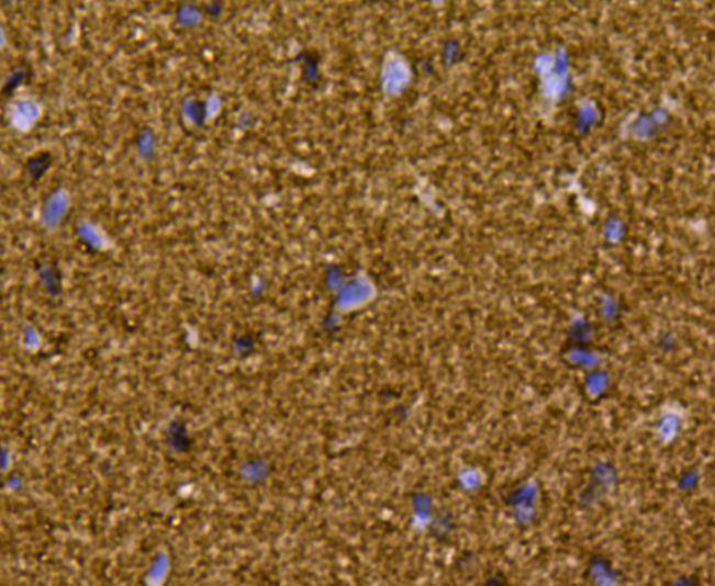
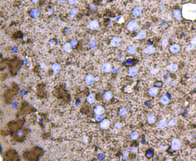
Fig6: Immunohistochemical analysis of paraffin-embedded rat brain tissue using anti-GAP43 antibody. Counter stained with hematoxylin.
Fig7: Immunohistochemical analysis of paraffin-embedded mouse brain tissue using anti-GAP43 antibody. Counter stained with hematoxylin.
- Background
-
References
- N-CAM modulates tumour-cell adhesion to matrix by inducing FGF-receptor signalling. Cavallaro U., Niedermeyer J., Fuxa M., Christofori G. Nat. Cell Biol. 3:650-657(2001)
- Acyl-protein thioesterase 2 catalyzes the deacylation of peripheral membrane-associated GAP-43. Tomatis V.M., Trenchi A., Gomez G.A., Daniotti J.L. PLoS ONE 5:E15045-E15045(2010)
Related Products / Services
Please note: All products are "FOR RESEARCH USE ONLY AND ARE NOT INTENDED FOR DIAGNOSTIC OR THERAPEUTIC USE"