
-
Product Name
Anti-FPR1 antibody
- Documents
-
Description
Rabbit polyclonal antibody to FPR1
-
Tested applications
WB, ICC, IHC-P, FC
-
Species reactivity
Human, Mouse
-
Alternative names
FPR antibody; FMLP antibody
-
Isotype
Rabbit IgG
-
Preparation
This antigen of this antibody was peptide
-
Clonality
Polyclonal
-
Formulation
Liquid, 1*PBS (pH7.4), 0.2% BSA, 40% Glycerol. Preservative: 0.05% Sodium Azide.
-
Storage instructions
Store at +4℃ after thawing. Aliquot store at -20℃ or -80℃. Avoid repeated freeze / thaw cycles.
-
Applications
WB: 1:500
ICC: 1:50-1:200
IHC-P: 1:50-1:100
FC: 1:50-1:100
-
Validations
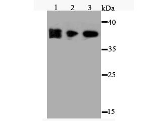
Fig1: Western blot analysis of FPR1 on different cell lysate using anti-FPR1 antibody at 1/500 dilution.; Positive control:; Lane 1: NCCIT Lane 2: MEF; Lane 3: HES
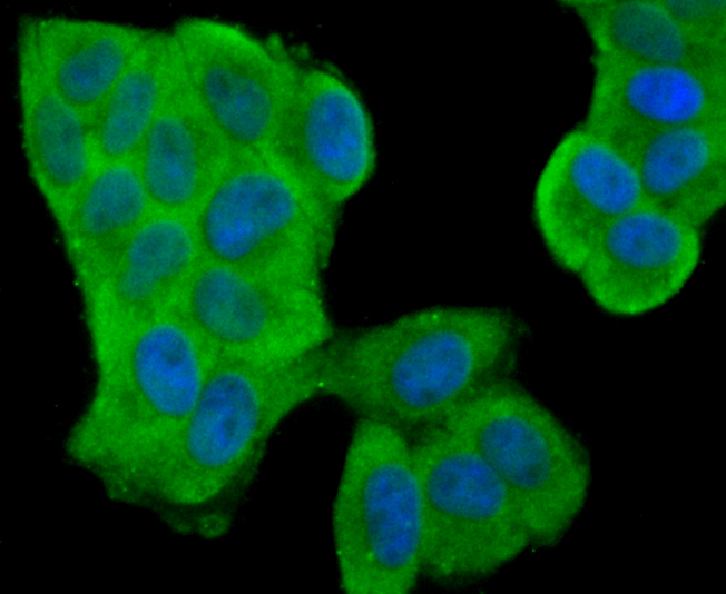
Fig2: ICC staining FPR1 in Hela cells (green). The nuclear counter stain is DAPI (blue). Cells were fixed in paraformaldehyde, permeabilised with 0.25% Triton X100/PBS.
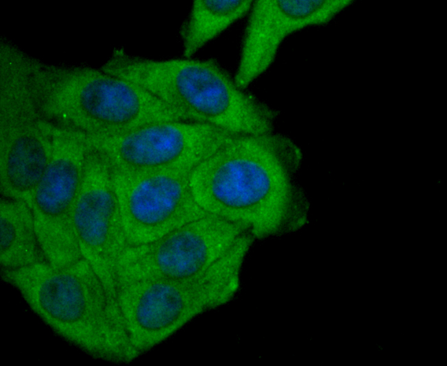
Fig3: ICC staining FPR1 in HepG2 cells (green). The nuclear counter stain is DAPI (blue). Cells were fixed in paraformaldehyde, permeabilised with 0.25% Triton X100/PBS.
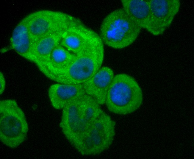
Fig4: ICC staining FPR1 in MCF-7 cells (green). The nuclear counter stain is DAPI (blue). Cells were fixed in paraformaldehyde, permeabilised with 0.25% Triton X100/PBS.
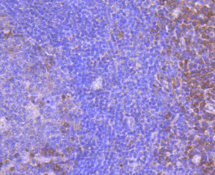
Fig5: Immunohistochemical analysis of paraffin-embedded human tonsil tissue using anti-FPR1 antibody. Counter stained with hematoxylin.
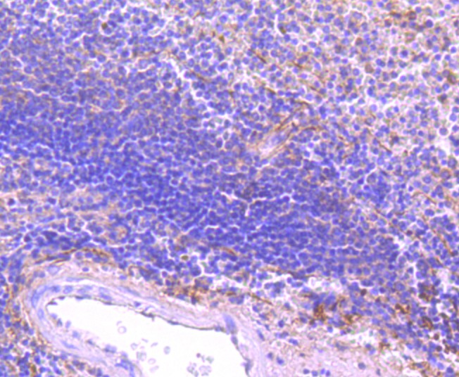
Fig6: Immunohistochemical analysis of paraffin-embedded human spleen tissue using anti-FPR1 antibody. Counter stained with hematoxylin.
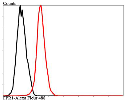
Fig7: Flow cytometric analysis of MCF-7 cells with FPR1 antibody at 1/100 dilution (red) compared with an unlabelled control (cells without incubation with primary antibody; black).
- Background
Related Products / Services
Please note: All products are "FOR RESEARCH USE ONLY AND ARE NOT INTENDED FOR DIAGNOSTIC OR THERAPEUTIC USE"