
-
Product Name
Anti-DOP1B antibody
- Documents
-
Description
Rabbit polyclonal antibody to DOP1B
-
Tested applications
WB, ICC, IHC-P, FC
-
Species reactivity
Human, Mouse
-
Alternative names
21orf5 antibody; DOPEY2 antibody; C21orf5 antibody
-
Isotype
Rabbit IgG
-
Preparation
This antigen of this antibody was synthetic peptide within human dopey2 aa 1030-1094.
-
Clonality
Polyclonal
-
Formulation
Liquid, 1*PBS (pH7.4), 0.2% BSA, 40% Glycerol. Preservative: 0.05% Sodium Azide.
-
Storage instructions
Store at +4℃ after thawing. Aliquot store at -20℃ or -80℃. Avoid repeated freeze / thaw cycles.
-
Applications
WB: 1:500-1:1,000
IHC-P: 1:50-1:200
ICC: 1:50-1:200
FC: 1:50-1:100
-
Validations
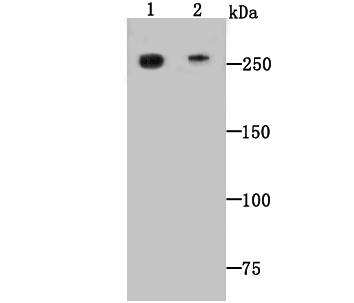
Fig1: Western blot analysis of Dopey-2 on mouse heart (1) and human heart (2) tissue lysate using anti-Dopey-2 antibody at 1/500 dilution.
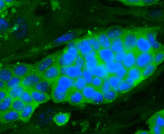
Fig2: ICC staining Dopey-2 in D3 cells (green). The nuclear counter stain is DAPI (blue). Cells were fixed in paraformaldehyde, permeabilised with 0.25% Triton X100/PBS.
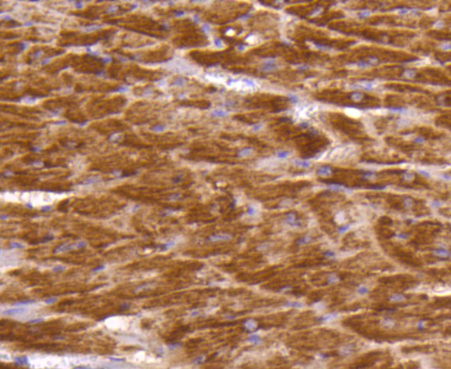
Fig3: Immunohistochemical analysis of formalin-fixed, paraffin-embedded mouse heart tissue labeling Dopey-2. Counterstained with Hematoxylin.
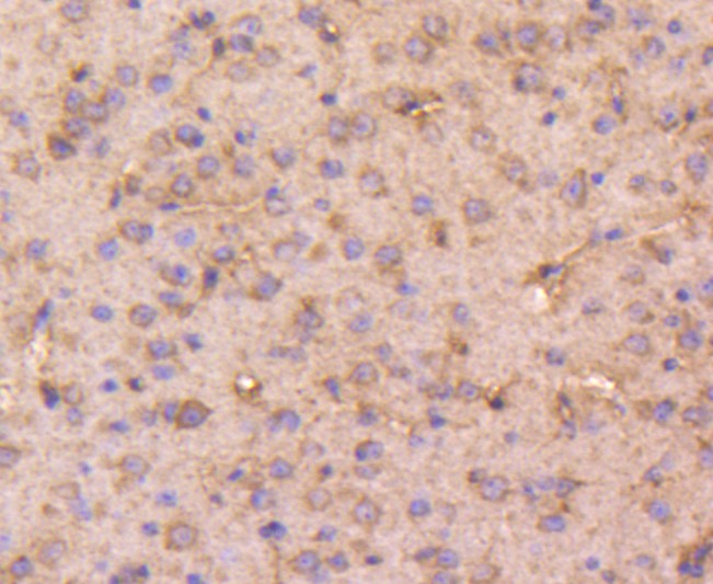
Fig4: Immunohistochemical analysis of formalin-fixed, paraffin-embedded mouse brain tissue labeling Dopey-2. Counterstained with Hematoxylin.
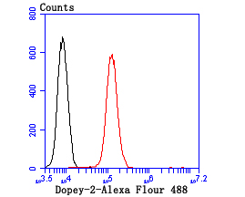
Fig5: Flow cytometric analysis of N2A cells with Dopey-2 antibody at 1/100 dilution (red) compared with an unlabelled control (cells without incubation with primary antibody; black).Alexa Fluor 488-conjugated goat anti-rabbit IgG was used as the secondary antibody.
- Background
-
References
- Rachidi M et al. A quantitative assessment of gene expression (QAGE) reveals differential overexpression of DOPEY2, a candidate gene for mental retardation, in Down syndrome brain regions. Int J Dev Neurosci 27(4):393-8 (2009).
- Swaminathan S et al. Analysis of copy number variation in Alzheimer's disease: the NIALOAD/ NCRAD Family Study. Curr Alzheimer Res 9(7):801-14 (2012).
Related Products / Services
Please note: All products are "FOR RESEARCH USE ONLY AND ARE NOT INTENDED FOR DIAGNOSTIC OR THERAPEUTIC USE"