-
Product Name
Anti-Cytokeratin 5 (9G3) Mouse antibody
- Documents
-
Description
Cytokeratin 5 (9G3) Mouse monoclonal antibody
-
Tested applications
IHC-P, ICC/IF
-
Species reactivity
Human
-
Isotype
Mouse IgG1
-
Preparation
Antigen: Purified recombinant fragment of CK5 expressed in E. Coli.
-
Clonality
Monoclonal
-
Formulation
Ascitic fluid containing 0.03% sodium azide.
-
Storage instructions
Store at 4°C short term. Store at -20°C long term. Avoid freeze / thaw cycle.
-
Applications
IHC: 1/200 - 1/1000
ICC: 1/200 - 1/1000
ELISA: 1/10000
-
Validations

Immunofluorescent analysis of HeLa cells fixed with -20℃ Methanol and using Cytokeratin (Pan) Mouse mAb (168518,dilution 1:500,green). DAPI was used to stain nucleus(blue).

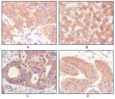
Immunohistochemical analysis of paraffin-embedded human lung squamous cell carcinoma (A),normal hepatocyte (B), colon adenocacinoma?, normal stomach tissue (D), showing cytoplasmic and membrane localization using CK mouse mAb with DAB staining.

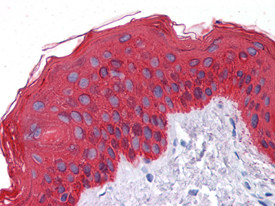
Immunohistochemical analysis of paraffin-embedded human Skin tissues using CK mouse mAb

Western blot analysis using CK mouse mAb against truncated CK5 recombinant protein.

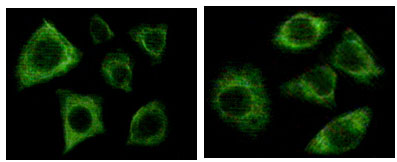
Immunofluorescence staining of methanol-fixed Eca-109 (left) and HepG2 (right) cells showing cytoplasmic localization.
-
Background
Swiss-Prot Acc.P13647.Biochemically, most members of the CK family fall into one of two classes, type I (acidic polypeptides) and type II (basic polypeptides). The type II cytokeratins consist of basic or neutral proteins which are arranged in pairs of heterotypic keratin chains coexpressed during differentiation of simple and stratified epithelial tissues. Cytokeratins comprise a diverse group of intermediate filament proteins (IFPs) that are expressed as pairs in both keratinized and non-keratinized epithelial tissue. Cytokeratins play a critical role in differentiation and tissue specialization and function to maintain the overall structural integrity of epithelial cells. Cytokeratins have been found to be useful markers of tissue differentiation which is directly applicable to the characterization of malignant tumors.
Related Products / Services
Please note: All products are "FOR RESEARCH USE ONLY AND ARE NOT INTENDED FOR DIAGNOSTIC OR THERAPEUTIC USE"
