
-
Product Name
Anti-CFH antibody
- Documents
-
Description
Rabbit monoclonal antibody to CFH
-
Tested applications
WB, ICC, IHC-P
-
Species reactivity
Human
-
Alternative names
FH antibody; HF antibody; HF1 antibody; HF2 antibody; HUS antibody; FHL1 antibody; AHUS1 antibody; AMBP1 antibody; ARMD4 antibody; ARMS1 antibody; CFHL3 antibody
-
Isotype
Rabbit IgG
-
Preparation
This antigen of this antibody was synthetic peptide within human factor h aa 150-200.
-
Clonality
Monoclonal
-
Formulation
Liquid, 1*TBS (pH7.4), 0.05% BSA, 40% Glycerol. Preservative: 0.05% Sodium Azide.
-
Storage instructions
Store at +4℃ after thawing. Aliquot store at -20℃ or -80℃. Avoid repeated freeze / thaw cycles.
-
Applications
WB: 1:500-1:2,000
ICC: 1:50-1:100
IHC-P: 1:50-1:100
-
Validations
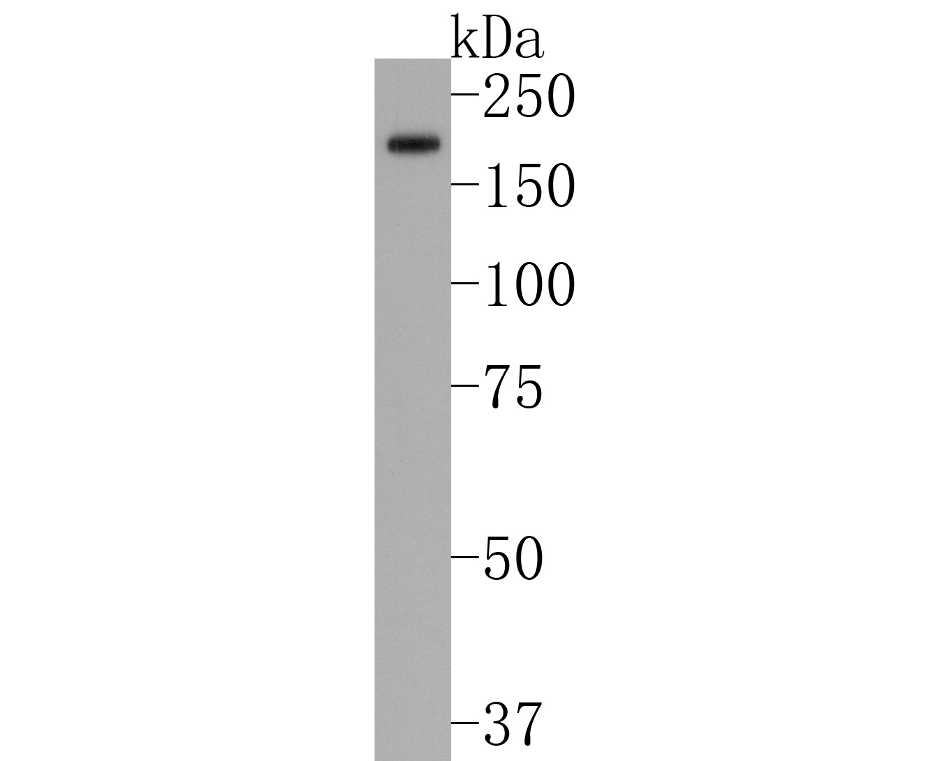
Fig1:; Western blot analysis of Factor H on human kidney tissue lysates. Proteins were transferred to a PVDF membrane and blocked with 5% BSA in PBS for 1 hour at room temperature. The primary antibody ( 1/500) was used in 5% BSA at room temperature for 2 hours. Goat Anti-Rabbit IgG - HRP Secondary Antibody (HA1001) at 1:200,000 dilution was used for 1 hour at room temperature.
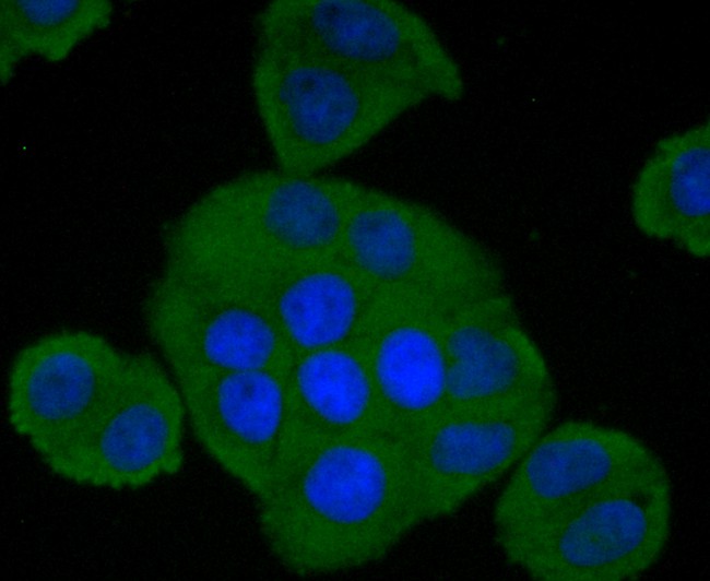
Fig2:; ICC staining of Factor H in Hela cells (green). Formalin fixed cells were permeabilized with 0.1% Triton X-100 in TBS for 10 minutes at room temperature and blocked with 1% Blocker BSA for 15 minutes at room temperature. Cells were probed with the primary antibody ( 1/50) for 1 hour at room temperature, washed with PBS. Alexa Fluor®488 Goat anti-Rabbit IgG was used as the secondary antibody at 1/1,000 dilution. The nuclear counter stain is DAPI (blue).
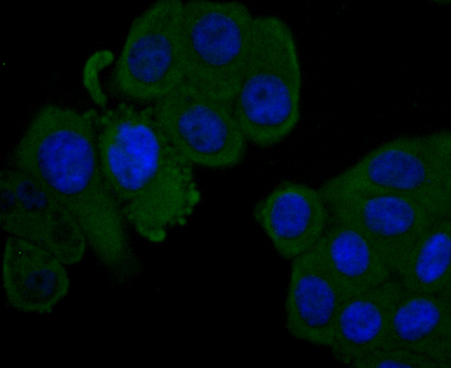
Fig3:; ICC staining of Factor H in HepG2 cells (green). Formalin fixed cells were permeabilized with 0.1% Triton X-100 in TBS for 10 minutes at room temperature and blocked with 1% Blocker BSA for 15 minutes at room temperature. Cells were probed with the primary antibody ( 1/50) for 1 hour at room temperature, washed with PBS. Alexa Fluor®488 Goat anti-Rabbit IgG was used as the secondary antibody at 1/1,000 dilution. The nuclear counter stain is DAPI (blue).
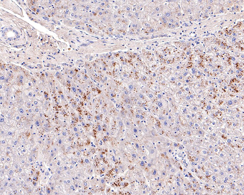
Fig4:; Immunohistochemical analysis of paraffin-embedded human liver tissue using anti-Factor H antibody. The section was pre-treated using heat mediated antigen retrieval with Tris-EDTA buffer (pH 8.0-8.4) for 20 minutes.The tissues were blocked in 5% BSA for 30 minutes at room temperature, washed with ddH; 2; O and PBS, and then probed with the primary antibody ( 1/100) for 30 minutes at room temperature. The detection was performed using an HRP conjugated compact polymer system. DAB was used as the chromogen. Tissues were counterstained with hematoxylin and mounted with DPX.
- Background
-
References
- Ermert D et al. Virulence of Group A Streptococci Is Enhanced by Human Complement Inhibitors. PLoS Pathog 11:e1005043 (2015).
- Segers FM et al. Complement alternative pathway activation in human nonalcoholic steatohepatitis. PLoS One 9:e110053 (2014).
Related Products / Services
Please note: All products are "FOR RESEARCH USE ONLY AND ARE NOT INTENDED FOR DIAGNOSTIC OR THERAPEUTIC USE"