
-
Product Name
Anti-CDC40 antibody
- Documents
-
Description
Rabbit monoclonal antibody to CDC40
-
Tested applications
WB, ICC/IF, IHC-P, FC
-
Species reactivity
Human, Mouse, Rat
-
Alternative names
EHB3 antibody; PCH15 antibody; PRP17 antibody; PRPF17 antibody
-
Isotype
Rabbit IgG
-
Preparation
This antigen of this antibody was recombinant protein
-
Clonality
Monoclonal
-
Formulation
Liquid, 1*TBS (pH7.4), 0.05% BSA, 40% Glycerol. Preservative: 0.05% Sodium Azide.
-
Storage instructions
Store at +4℃ after thawing. Aliquot store at -20℃ or -80℃. Avoid repeated freeze / thaw cycles.
-
Applications
WB: 1:1,000-1:5,000
ICC/IF: 1:50-1:200
IHC-P: 1:50-1:200
FC: 1:50-1:100
-
Validations
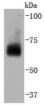
Fig1: Western blot analysis of CDC40 on Hela cells lysates using anti-CDC40 antibody at 1/2,000 dilution.
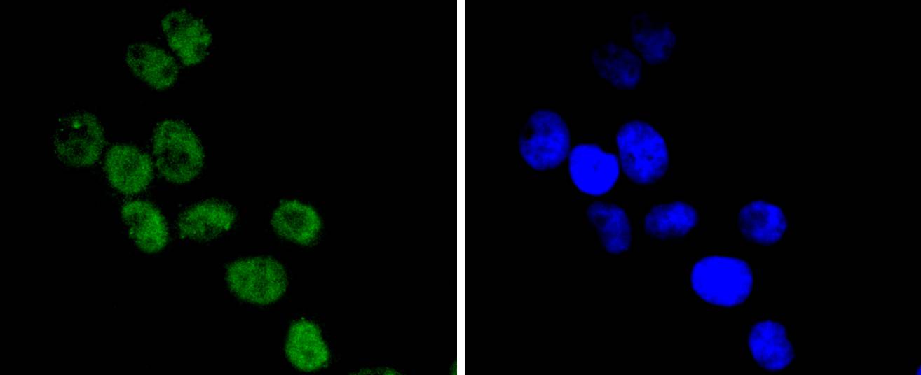
Fig2: ICC staining CDC40 in Hela cells (green). The nuclear counter stain is DAPI (blue). Cells were fixed in paraformaldehyde, permeabilised with 0.25% Triton X100/PBS.
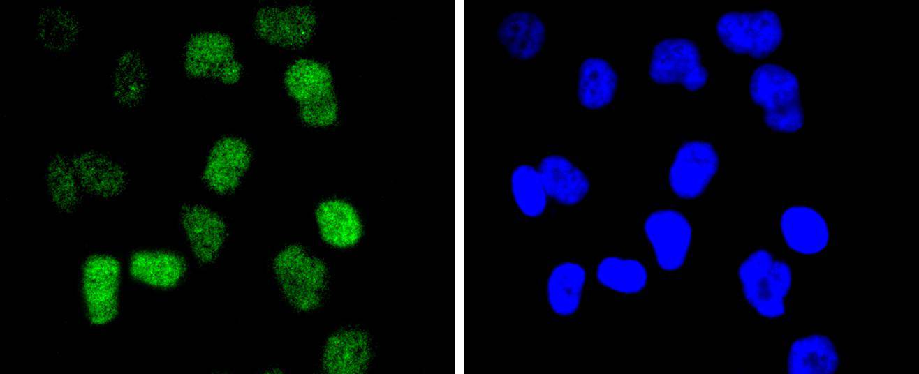
Fig3: ICC staining CDC40 in HUVEC cells (green). The nuclear counter stain is DAPI (blue). Cells were fixed in paraformaldehyde, permeabilised with 0.25% Triton X100/PBS.
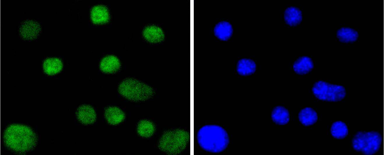
Fig4: ICC staining CDC40 in SHG-44 cells (green). The nuclear counter stain is DAPI (blue). Cells were fixed in paraformaldehyde, permeabilised with 0.25% Triton X100/PBS.
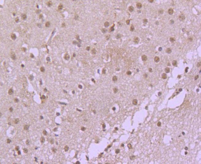
Fig5: Immunohistochemical analysis of paraffin-embedded rat brain tissue using anti-CDC40 antibody. Counter stained with hematoxylin.
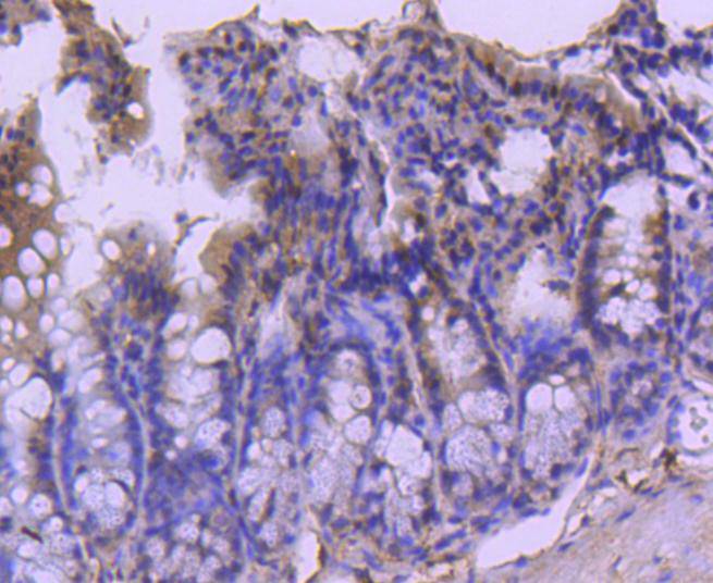
Fig6: Immunohistochemical analysis of paraffin-embedded mouse colon tissue using anti-CDC40 antibody. Counter stained with hematoxylin.
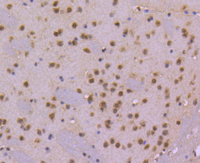
Fig7: Immunohistochemical analysis of paraffin-embedded mouse brain tissue using anti-CDC40 antibody. Counter stained with hematoxylin.
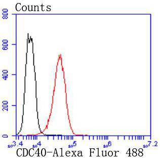
Fig8: Flow cytometric analysis of K562 cells with CDC40 antibody at 1/50 dilution (red) compared with an unlabelled control (cells without incubation with primary antibody; black). Alexa Fluor 488-conjugated goat anti rabbit IgG was used as the secondary
- Background
-
References
- Olsen JV, et al. 2006. Global, in vivo, and site-specific phosphorylation dynamics in signaling networks. Cell. 127 (3): 635–48.
- Barrios-Rodiles M, et al. 2005. High-throughput mapping of a dynamic signaling network in mammalian cells. Science. 307 (5715): 1621–5.
Related Products / Services
Please note: All products are "FOR RESEARCH USE ONLY AND ARE NOT INTENDED FOR DIAGNOSTIC OR THERAPEUTIC USE"