
-
Product Name
Anti-CBL (6C5) Mouse antibody
- Documents
-
Description
CBL (6C5) Mouse monoclonal antibody
-
Tested applications
WB, IHC-P, ICC/IF, FC, IP
-
Species reactivity
Human, Mouse, Rat
-
Isotype
Mouse IgG1
-
Preparation
Antigen: Purified recombinant fragment of human C-CBL expressed in E. Coli.
-
Clonality
Monoclonal
-
Formulation
Ascitic fluid containing 0.03% sodium azide.
-
Storage instructions
Store at 4°C short term. Store at -20°C long term. Avoid freeze / thaw cycle.
-
Applications
WB: 1/500 - 1/2000
IHC: 1/200 - 1/1000
ICC: 1/200 - 1/1000
FC: 1/200 - 1/400
ELISA: 1/10000
-
Validations
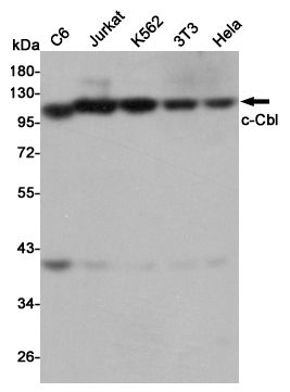
Western blot detection of c-Cbl in C6,Jurkat,K562,3T3 and Hela cell lysates using c-Cbl mouse mAb (1:1000 diluted).Predicted band size:120KDa.Observed band size:120KDa.
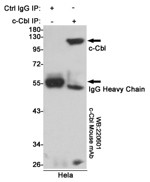
Immunoprecipitation analysis of Hela cell lysates using c-Cbl mouse mAb.
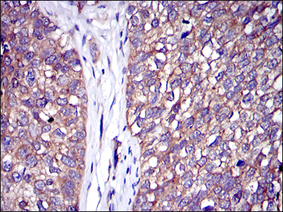
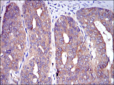
Immunohistochemical analysis of paraffin-embedded bladder cancer tissues using C-CBL mouse mAb with DAB staining.
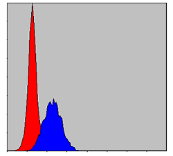
Flow cytometric analysis of MCF-7 cells using C-CBL mouse mAb (blue) and negative control (red).
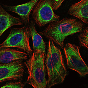
Immunofluorescence analysis of Hela cells using C-CBL mouse mAb (green). Blue
Immunohistochemical analysis of paraffin-embedded ovarian cancer tissues using C-CBL mouse mAb with DAB staining.
-
Background
Swiss-Prot Acc.P22681.The cbl oncogene was first identified as part of a transforming retrovirus which induces mouse pre-B and pro-B cell lymphomas. As an adaptor protein for receptor protein-tyrosine kinases, it positively regulates receptor protein-tyrosine kinase ubiquitination in a manner dependent upon its variant SH2 and RING finger domains. Ubiquitination of receptor protein-tyrosine kinases terminates signaling by marking active receptors for degradation.
Related Products / Services
Please note: All products are "FOR RESEARCH USE ONLY AND ARE NOT INTENDED FOR DIAGNOSTIC OR THERAPEUTIC USE"