
-
Product Name
Anti-ATRAID antibody
- Documents
-
Description
Rabbit polyclonal antibody to ATRAID
-
Tested applications
WB, ICC, IHC-P, FC
-
Species reactivity
Human, Mouse, Rat
-
Alternative names
p18 antibody; APR3 antibody; APR-3 antibody; APR--3 antibody; PRO240 antibody; C2orf28 antibody; HSPC013 antibody
-
Isotype
Rabbit IgG
-
Preparation
This antigen of this antibody was synthetic peptide corresponding to mouse apr3 c terminal.
-
Clonality
Polyclonal
-
Formulation
Liquid, 1*PBS (pH7.4), 0.2% BSA, 50% Glycerol. Preservative: 0.05% Sodium Azide.
-
Storage instructions
Store at +4℃ after thawing. Aliquot store at -20℃. Avoid repeated freeze / thaw cycles.
-
Applications
WB: 1:500-2,000
ICC: 1:50-1:200
IHC-P: 1:50-1:200
FC: 1:50-1:100
-
Validations
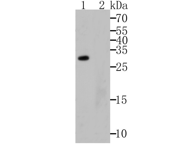
Fig1: Western blot analysis of APR3 on human skin tissue lysates using anti-APR3 antibody.; Lane 1: Anti-APR3 antibody (1/500).; Lane 2: Anti-APR3 antibody, pre-incubated with the immunizaiton peptide.
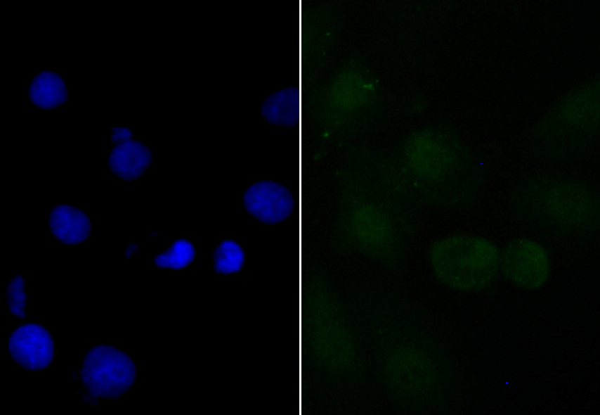
Fig2: ICC staining APR3 in A549 cells (green). The nuclear counter stain is DAPI (blue). Cells were fixed in paraformaldehyde, permeabilised with 0.25% Triton X100/PBS.
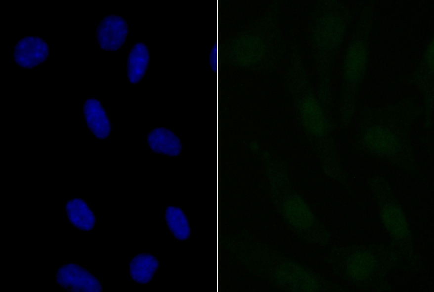
Fig3: ICC staining APR3 in SH-SY-5Y cells (green). The nuclear counter stain is DAPI (blue). Cells were fixed in paraformaldehyde, permeabilised with 0.25% Triton X100/PBS.
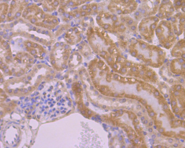
Fig4: Immunohistochemical analysis of paraffin-embedded mouse liver tissue using anti-APR3 antibody. Counter stained with hematoxylin.
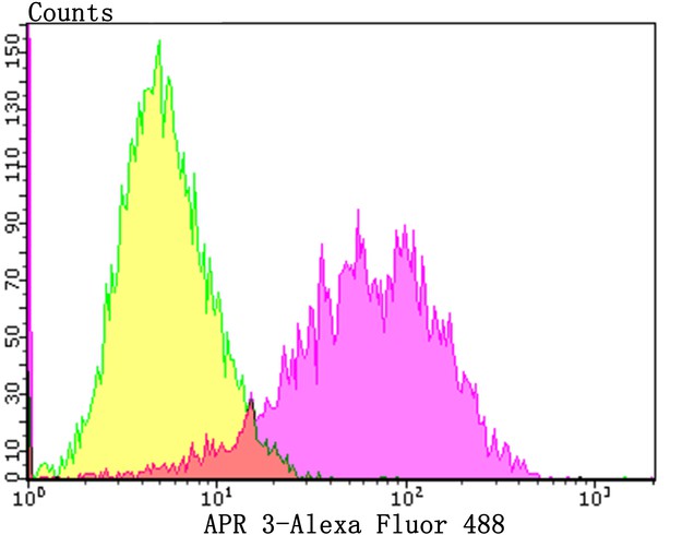
Fig5: Flow cytometric analysis of MCF-7 cells with APR3 antibody at 1/100 dilution (Pink purple) compared with an unlabelled control (cells without incubation with primary antibody; Yellow). Alexa Fluor 488-conjugated goat anti-rabbit IgG was used as the secondary antibody.
- Background
-
References
- Zou X et al. NELL-1 binds to APR3 affecting human osteoblast proliferation and differentiation. FEBS Lett. 585:2410-2418 (2011).
- Yu F et al. Apoptosis related protein 3, an ATRA-upregulated membrane protein arrests the cell cycle at G1/S phase by decreasing the expression of cyclin D1. Biochem. Biophys. Res. Commun. 358:1041-1046 (2007).
Related Products / Services
Please note: All products are "FOR RESEARCH USE ONLY AND ARE NOT INTENDED FOR DIAGNOSTIC OR THERAPEUTIC USE"