

-
Product Name
Anti-ASIC1 antibody
- Documents
-
Description
Rabbit polyclonal antibody to ASIC1
-
Tested applications
WB, IHC-P, FC
-
Species reactivity
Human, Mouse, Rat
-
Alternative names
ASIC antibody; ACCN2 antibody; BNaC2 antibody
-
Isotype
Rabbit IgG
-
Preparation
This antigen of this antibody was synthetic peptide within human accn2 aa 460-500.
-
Clonality
Polyclonal
-
Formulation
Liquid, 1*PBS (pH7.4), 0.2% BSA, 50% Glycerol. Preservative: 0.05% Sodium Azide.
-
Storage instructions
Store at +4℃ after thawing. Aliquot store at -20℃. Avoid repeated freeze / thaw cycles.
-
Applications
WB: 1:500-1:2,000
IHC-P: 1:50-1:200
FC: 1:50-1:100
-
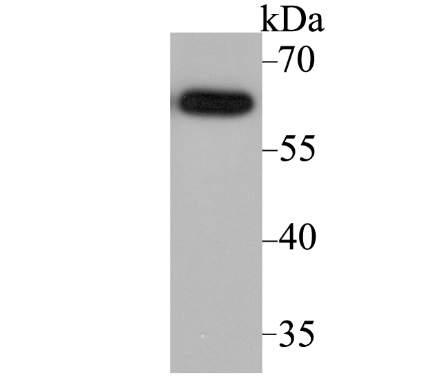
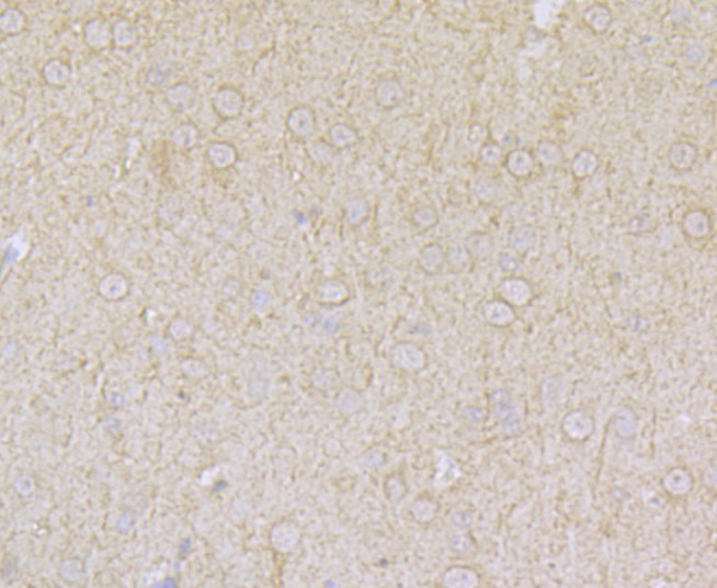
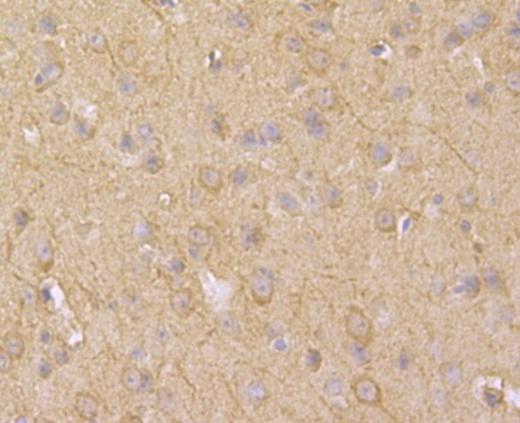
Validations
Fig1: Western blot analysis of ACCN2 on SH-SY-5Y cell lysate using anti-ACCN2 antibody at 1/2,000 dilution.
Fig2: Immunohistochemical analysis of paraffin-embedded rat brain tissue using anti-ACCN2 antibody. Counter stained with hematoxylin.
Fig3: Immunohistochemical analysis of paraffin-embedded mouse brain tissue using anti-ACCN2 antibody. Counter stained with hematoxylin.
Fig4: Flow cytometric analysis of SH-SY-5Y cells with ACCN2 antibody at 1/100 dilution (fuchsia) compared with an unlabelled control (cells without incubation with primary antibody; yellow). Alexa Fluor 488-conjugated goat anti-rabbit IgG was used as the secondary antibody.
- Background
-
References
- Dawson R J et al. Structure of the acid-sensing ion channel 1 in complex with the gating modifier Psalmotoxin 1. Nat Commun 3:936-943 (2012).
- Sherwood T et al. Identification of protein domains that control proton and calcium sensitivity of ASIC1a. J Biol Chem 284:27899-27907 (2009).
Related Products / Services
Please note: All products are "FOR RESEARCH USE ONLY AND ARE NOT INTENDED FOR DIAGNOSTIC OR THERAPEUTIC USE"