
-
Product Name
Anti-ABCF1 antibody
- Documents
-
Description
Rabbit monoclonal antibody to ABCF1
-
Tested applications
WB, ICC/IF, IHC-P
-
Species reactivity
Human, Rat
-
Alternative names
ABC27 antibody; ABC50 antibody
-
Isotype
Rabbit IgG
-
Preparation
This antigen of this antibody was recombinant protein
-
Clonality
Monoclonal
-
Formulation
Liquid, 1*TBS (pH7.4), 0.05% BSA, 40% Glycerol. Preservative: 0.05% Sodium Azide.
-
Storage instructions
Store at +4℃ after thawing. Aliquot store at -20℃ or -80℃. Avoid repeated freeze / thaw cycles.
-
Applications
WB: 1:1,000-1:2,000
ICC/IF: 1:100-1:500
IHC-P: 1:50-1:200
-
Validations
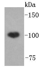
Fig1: Western blot analysis of ABCF1 on Hela lysates using anti-ABCF1 antibody at 1/1,000 dilution.
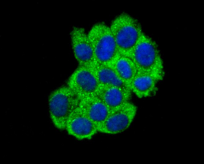
Fig2: ICC staining ABCF1 in HepG2 cells (green). The nuclear counter stain is DAPI (blue). Cells were fixed in paraformaldehyde, permeabilised with 0.25% Triton X100/PBS.
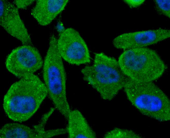
Fig3: ICC staining ABCF1 in PC-3M cells (green). The nuclear counter stain is DAPI (blue). Cells were fixed in paraformaldehyde, permeabilised with 0.25% Triton X100/PBS.
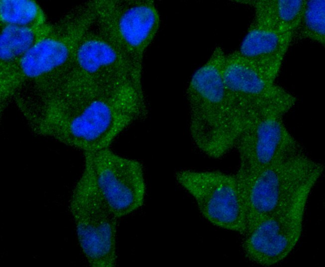
Fig4: ICC staining ABCF1 in Hela cells (green). The nuclear counter stain is DAPI (blue). Cells were fixed in paraformaldehyde, permeabilised with 0.25% Triton X100/PBS.
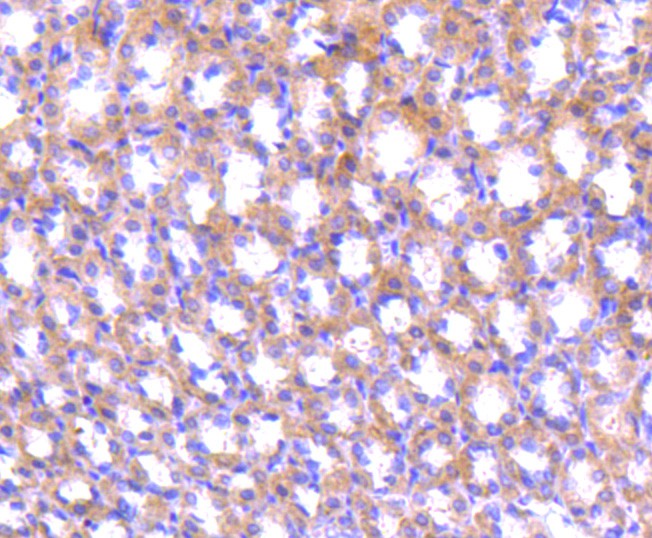
Fig5: Immunohistochemical analysis of paraffin-embedded rat stomach tissue using anti-ABCF1 antibody. Counter stained with hematoxylin.
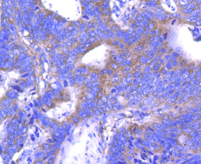
Fig6: Immunohistochemical analysis of paraffin-embedded human colon cancer tissue using anti-ABCF1 antibody. Counter stained with hematoxylin.
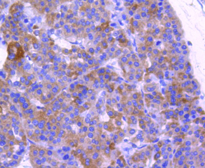
Fig7: Immunohistochemical analysis of paraffin-embedded human liver cancer tissue using anti-ABCF1 antibody. Counter stained with hematoxylin.
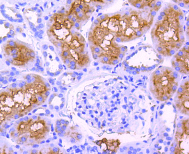
Fig8: Immunohistochemical analysis of paraffin-embedded human kidney tissue using anti-ABCF1 antibody. Counter stained with hematoxylin.
- Background
-
References
- Pechenino AS et al. Impact of aging vs. estrogen loss on cardiac gene expression: estrogen replacement and inflammation. Physiol Genomics 43:1065-73 (2011).
- DU, P. et al. Metastasis Suppressor-1, MTSS1, Acts as a Putative Tumour Suppressor in Human Bladder Cancer. Anticancer Res 31: 3205-3212 (2011).
Related Products / Services
Please note: All products are "FOR RESEARCH USE ONLY AND ARE NOT INTENDED FOR DIAGNOSTIC OR THERAPEUTIC USE"