
-
Product Name
Aconitase 2 antibody
- Documents
-
Description
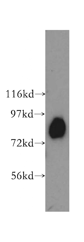
Aconitase 2 Rabbit Polyclonal antibody. Positive WB detected in human skeletal muscle tissue, HeLa cells, human heart tissue, human liver tissue, mouse heart tissue. Positive IHC detected in human lung cancer tissue, human prostate cancer tissue. Observed molecular weight by Western-blot: 85kd
-
Tested applications
ELISA, WB, IHC
-
Species reactivity
Human,Mouse,Rat; other species not tested.
-
Alternative names
ACO2 antibody; Aconitase antibody; aconitase 2 antibody; aconitase 2 antibody; mitOChondrial antibody; ACONM antibody; Citrate hydro lyase antibody
-
Isotype
Rabbit IgG
-
Preparation
This antibody was obtained by immunization of Aconitase 2 recombinant protein (Accession Number: NM_001098). Purification method: Antigen affinity purified.
-
Clonality
Polyclonal
-
Formulation
PBS with 0.1% sodium azide and 50% glycerol pH 7.3.
-
Storage instructions
Store at -20℃. DO NOT ALIQUOT
-
Applications
Recommended Dilution:
WB: 1:500-1:5000
IHC: 1:20-1:200
-
Validations
human skeletal muscle tissue were subjected to SDS PAGE followed by western blot with Catalog No:107742(ACO2 antibody) at dilution of 1:400
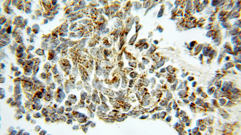
Immunohistochemical of paraffin-embedded human lung cancer using Catalog No:107742(ACO2 antibody) at dilution of 1:50 (under 10x lens)
-
Background
ACO2(aconitate hydratase, mitochondrial) is also named as citrate hydro-lyase and belongs to the aconitase/IPM isomerase family. It plays a key function in cellular energy production, and loss of its activity has a major impact on cellular and organismal survival. Western blot shows two bands of 83 kDa and 40 kDa. The 40 kDa fragment decreases with age and oxidative stress, whereas other fragmentation products with molecular weights between 40 and 83 kDa increased with age and MnSOD(mitochondrial manganese superoxide dismutase) deficiency(PMID:12459471). Defects in ACO2 are the cause of infantile cerebellar-retinal degeneration (ICRD).
-
References
- Cai Z, Zhao JS, Li JJ. A combined proteomics and metabolomics profiling of gastric cardia cancer reveals characteristic dysregulations in glucose metabolism. Molecular & cellular proteomics : MCP. 9(12):2617-28. 2010.
- Shin DI, Oh YJ. Tumor Necrosis Factor-Associated Protein 1 (TRAP1) is Released from the Mitochondria Following 6-hydroxydopamine Treatment. Experimental neurobiology. 23(1):65-76. 2014.
Related Products / Services
Please note: All products are "FOR RESEARCH USE ONLY AND ARE NOT INTENDED FOR DIAGNOSTIC OR THERAPEUTIC USE"