
-
Product Name
14-3-3 antibody
- Documents
-
Description
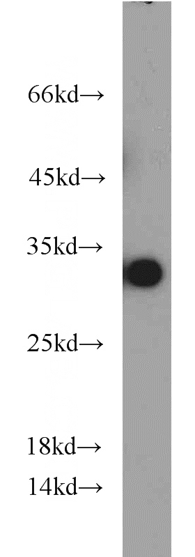
14-3-3 Mouse Monoclonal antibody. Positive FC detected in HeLa cells. Positive IF detected in HepG2 cells. Positive IHC detected in rat brain tissue. Positive WB detected in human brain tissue, HeLa cells, HepG2 cells. Observed molecular weight by Western-blot: 31 kDa,28 kDa
-
Tested applications
ELISA, WB, IHC, IF, FC
-
Species reactivity
Human, Rat; other species not tested.
-
Alternative names
14 3 3 antibody; 14 3 3 protein T cell antibody; 14 3 3 protein tau antibody; 14 3 3 protein theta antibody; 14-3-3 tau antibody; HS1 antibody; Protein HS1 antibody; YWHAQ antibody
-
Isotype
Mouse IgG1
-
Preparation
This antibody was obtained by immunization of 14-3-3 recombinant protein (Accession Number: NM_006826). Purification method: Protein G purified.
-
Clonality
Monoclonal
-
Formulation
PBS with 0.02% sodium azide and 50% glycerol pH 7.3.
-
Storage instructions
Store at -20℃. DO NOT ALIQUOT
-
Applications
Recommended Dilution:
WB: 1:500-1:5000
IHC: 1:20-1:200
IF: 1:20-1:200
-
Validations
human brain tissue were subjected to SDS PAGE followed by western blot with Catalog No:107550(14-3-3 antibody) at dilution of 1:1000
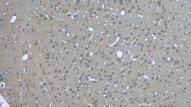
Immunohistochemistry of paraffin-embedded rat brain tissue slide using Catalog No:107550(14-3-3 Antibody) at dilution of 1:50 (under 10x lens)
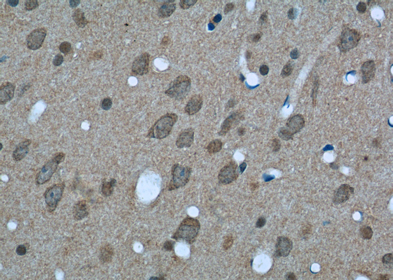
Immunohistochemistry of paraffin-embedded rat brain tissue slide using Catalog No:107550(14-3-3 Antibody) at dilution of 1:50 (under 40x lens)
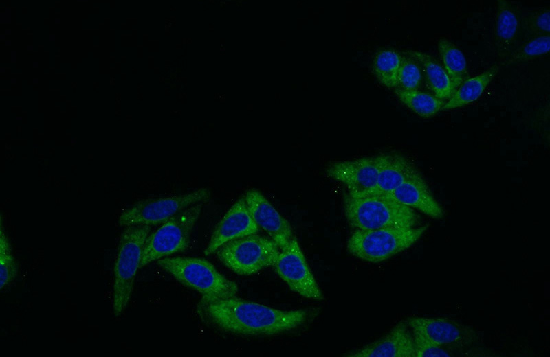
Immunofluorescent analysis of HepG2 cells using Catalog No:107550(14-3-3 Antibody) at dilution of 1:50 and Alexa Fluor 488-congugated AffiniPure Goat Anti-Mouse IgG(H+L)
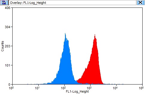
1X10^6 HeLa cells were stained with 0.2ug 14-3-3 antibody (Catalog No:107550, red) and control antibody (blue). Fixed with 90% MeOH blocked with 3% BSA (30 min). Alexa Fluor 488-congugated AffiniPure Goat Anti-Mouse IgG(H+L) with dilution 1:1500.
-
Background
14-3-3 proteins interact with a wide spectrum of proteins and possess diverse functions. Mammals express seven distinct 14-3-3 isoforms (gamma, epsilon, beta, zeta, sigma, theta, tau) that form multiple homo- and hetero- dimmers. 14-3-3 proteins display the highest expression levels in the brain, and have been implicated in several neurodegenerative diseases, including Alzheimer's disease and amyotrophic lateral sclerosis. 66061-1-Ig recognizes 14-3-3 tau (YWHAQ), eta (YWHAH), and zeta (YWHAZ), but not epsilon (YWHAE).
Related Products / Services
Please note: All products are "FOR RESEARCH USE ONLY AND ARE NOT INTENDED FOR DIAGNOSTIC OR THERAPEUTIC USE"