
-
Product Name
TGF-beta 1 antibody
- Documents
-
Description
TGF-beta 1 Rabbit Polyclonal antibody. Positive WB detected in MCF-7 cells, K-562 cells, mouse brain tissue, mouse uterus tissue, Raji cells. Positive IHC detected in human ovary tissue, human breast cancer tissue, human kidney tissue, human liver cancer tissue, human liver tissue, human lung cancer tissue, human lung tissue, human placenta tissue, human skin tissue, human spleen tissue, human testis tissue, rat ovary tissue. Observed molecular weight by Western-blot: 25 kDa
-
Tested applications
ELISA, WB, IHC
-
Species reactivity
Human,Mouse,Rat; other species not tested.
-
Alternative names
CED antibody; DPD1 antibody; LAP antibody; TGF beta 1 antibody; TGFB antibody; TGFB1 antibody; TGFbeta antibody; TGF-beta1 antibody; TGF-β1 antibody
-
Isotype
Rabbit IgG
-
Preparation
This antibody was obtained by immunization of Peptide (Accession Number: NM_000660). Purification method: Antigen affinity purified.
-
Clonality
Polyclonal
-
Formulation
PBS with 0.1% sodium azide and 50% glycerol pH 7.3.
-
Storage instructions
Store at -20℃. DO NOT ALIQUOT
-
Applications
Recommended Dilution:
WB: 1:200-1:2000
IHC: 1:50-1:500
-
Validations
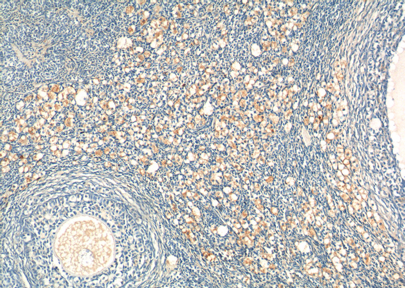
Immunohistochemistry of paraffin-embedded human ovary tissue slide using Catalog No:116019(TGFB1 Antibody) at dilution of 1:200 (under 10x lens).
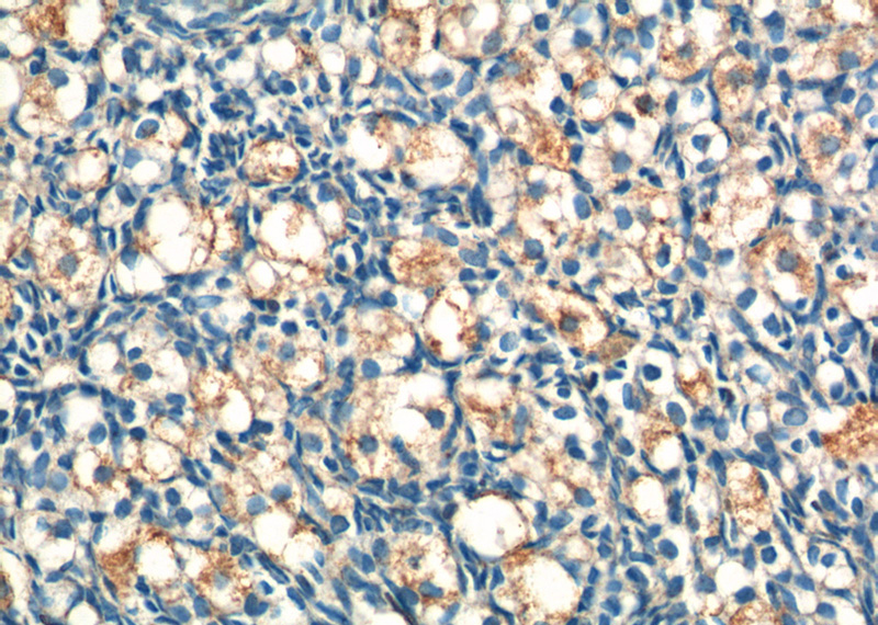
Immunohistochemistry of paraffin-embedded human ovary tissue slide using Catalog No:116019(TGFB1 Antibody) at dilution of 1:200 (under 40x lens).
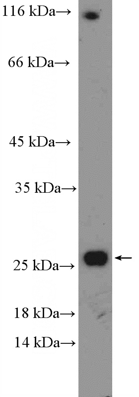
MCF-7 cells were subjected to SDS PAGE followed by western blot with Catalog No:116019(TGFB1 Antibody) at dilution of 1:600
-
Background
TGFB, also named as LAP and TGFB1, is a multifunctional peptide that controls proliferation, differentiation, and other functions in many cell types. TGFB acts synergistically with TGFA in inducing transformation. It also acts as a negative autocrine growth factor. Dysregulation of TGFB activation and signaling may result in apoptosis. Many cells synthesize TGFB and almost all of them have specific receptors for it. TGFB positively and negatively regulates many other growth factors. It plays an important role in bone remodeling as it is a potent stimulator of osteoblastic bone formation, causing chemotaxis, proliferation and differentiation in committed osteoblasts. It is highly expressed in bone. Mutation of TGFB are the cause of Camurati-Engelmann disease (CED) which known as progressive diaphyseal dysplasia 1 (DPD1).
-
References
- Zhang Q, Xiao X, Li M. Attenuating effect of Fufang Xueshuantong Capsule on kidney function in diabetic nephropathy model. Journal of natural medicines. 67(1):86-97. 2013.
- Li G, Li Y, Liu S. Gremlin aggravates hyperglycemia-induced podocyte injury by a TGFβ/smad dependent signaling pathway. Journal of cellular biochemistry. 114(9):2101-13. 2013.
- Li W, Cui M, Wei Y, Kong X, Tang L, Xu D. Inhibition of the expression of TGF-β1 and CTGF in human mesangial cells by exendin-4, a glucagon-like peptide-1 receptor agonist. Cellular physiology and biochemistry : international journal of experimental cellular physiology, biochemistry, and pharmacology. 30(3):749-57. 2012.
- Ding ZY, Jin GN, Liang HF. Transforming growth factor β induces expression of connective tissue growth factor in hepatic progenitor cells through Smad independent signaling. Cellular signalling. 25(10):1981-92. 2013.
- Wei J, Shi Y, Hou Y. Knockdown of thioredoxin-interacting protein ameliorates high glucose-induced epithelial to mesenchymal transition in renal tubular epithelial cells. Cellular signalling. 25(12):2788-96. 2013.
- Jia M, Hu J, Li W. Trps1 is associated with the multidrug resistance of osteosarcoma by regulating MDR1 gene expression. FEBS letters. 588(5):801-10. 2014.
- Qu Y, Ray PS, Li J. High levels of secreted frizzled-related protein 1 correlate with poor prognosis and promote tumourigenesis in gastric cancer. European journal of cancer (Oxford, England : 1990). 49(17):3718-28. 2013.
- Azzimonti B, Zavattaro E, Provasi M. Intense Foxp3+ CD25+ regulatory T-cell infiltration is associated with high-grade cutaneous squamous cell carcinoma and counterbalanced by CD8+/Foxp3+ CD25+ ratio. The British journal of dermatology. 172(1):64-73. 2015.
Related Products / Services
Please note: All products are "FOR RESEARCH USE ONLY AND ARE NOT INTENDED FOR DIAGNOSTIC OR THERAPEUTIC USE"