
-
Product Name
Anti-RAB6B antibody
- Documents
-
Description
Mouse monoclonal antibody to RAB6B
-
Tested applications
WB, ICC, IHC-P, FC
-
Species reactivity
Human, Rat
-
Isotype
Mouse IgG1
-
Preparation
This antigen of this antibody was recombinant protein
-
Clonality
Monoclonal
-
Formulation
Liquid, 1*TBS (pH7.4), 1%BSA, 40%Glycerol. Preservative: 0.05% Sodium Azide.
-
Storage instructions
Store at +4℃ after thawing. Aliquot store at -20℃ or -80℃. Avoid repeated freeze / thaw cycles.
-
Applications
WB: 1:500-1:2,000
ICC: 1:50-1:200
IHC-P: 1:50-1:200
FC: 1:50-1:200
-
Validations
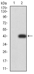
Fig1: Western blot analysis of Rab6b on human Rab6b recombinant protein using anti-Rab6b antibody at 1/1,000 dilution.
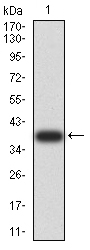
Fig2: Western blot analysis of Rab6b on HEK293 (1) and Rab6b-hIgGFc transfected HEK293 (2) cell lysate using anti-Rab6b antibody at 1/1,000 dilution.
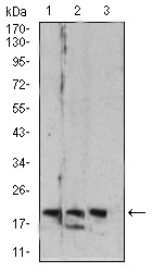
Fig3: Western blot analysis of Rab6b on different cell lysate using anti-Rab6b antibody at 1/1,000 dilution.; Positive control:; Lane 1: C6; Lane 2: HT-29; Lane 3: PC-12
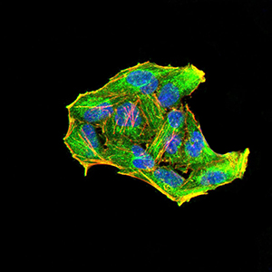
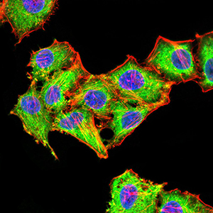
Fig4: ICC staining Rab6b (green) and Actin filaments (red) in Hela cells. The nuclear counter stain is DAPI (blue). Cells were fixed in paraformaldehyde, permeabilised with 0.25% Triton X100/PBS.
Fig5: ICC staining Rab6b (green) and Actin filaments (red) in HepG2 cells. The nuclear counter stain is DAPI (blue). Cells were fixed in paraformaldehyde, permeabilised with 0.25% Triton X100/PBS.
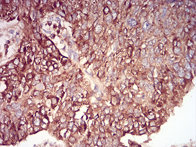
Fig6: Immunohistochemical analysis of paraffin-embedded human lung cancer tissue using anti-Rab6b antibody. Counter stained with hematoxylin.
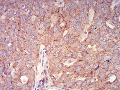
Fig7: Immunohistochemical analysis of paraffin-embedded human ovarian cancer tissue using anti-Rab6b antibody. Counter stained with hematoxylin.
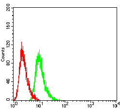
Fig8: Flow cytometric analysis of Hela cells with Rab6b antibody at 1/100 dilution (green) compared with an unlabelled control (cells without incubation with primary antibody; red).
- Background
-
References
- Petrosyan A et al. Restoration of compact Golgi morphology in advanced prostate cancer enhances susceptibility to galectin-1-induced apoptosis by modifying mucin O-glycan synthesis. Mol Cancer Res 12:1704-16 (2014).
- Petrosyan A & Cheng PW A non-enzymatic function of Golgi glycosyltransferases: mediation of Golgi fragmentation by interaction with non-muscle myosin IIA. Glycobiology 23:690-708 (2013).
Related Products / Services
Please note: All products are "FOR RESEARCH USE ONLY AND ARE NOT INTENDED FOR DIAGNOSTIC OR THERAPEUTIC USE"